Fraud Blocker Skip to main content

Macrophage Collection

Macrophages, the mighty defenders of our immune system, play a crucial role in protecting us from harmful invaders

Background imageMacrophage Collection: Macrophage engulfing TB bacteria, SEM

Macrophage engulfing TB bacteria, SEM
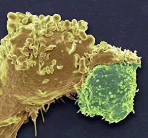
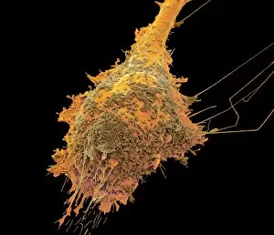
Macrophage engulfing TB bacteria. Coloured scanning electron micrograph (SEM) of a macrophage white blood cell (red) engulfing a tuberculosis (Mycobacterium tuberculosis) bacterium (yellow)

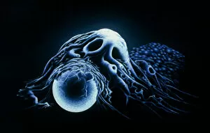
Background imageMacrophage Collection: Dendritic cells, artwork

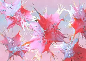
Dendritic cells, artwork. Dendritic cells, a type of white blood cell, are called antigen presenting cells (APCs) and are found in the bodys tissues

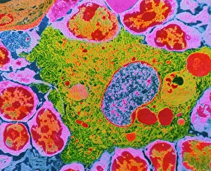
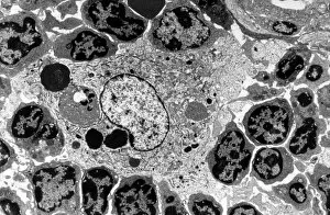
Background imageMacrophage Collection: TEM of human macrophage

TEM of human macrophage

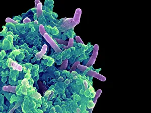
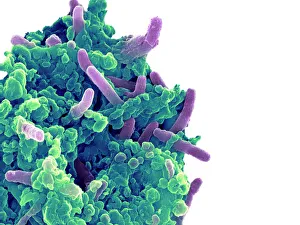
Background imageMacrophage Collection: Bacteria infecting a macrophage, SEM

Bacteria infecting a macrophage, SEM
Bacteria infecting a macrophage. Coloured scanning electron micrograph (SEM) of Mycobacterium tuberculosis bacteria (purple) infecting a macrophage white blood cell

Background imageMacrophage Collection: Activated macrophage, SEM C015 / 6375

Activated macrophage, SEM C015 / 6375
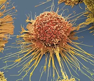
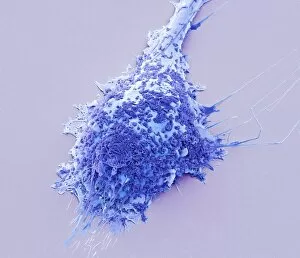
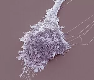
Activated macrophage. Coloured scanning electron micrograph (SEM) of an active macrophage white blood cell. Macrophages are cells of the bodys immune system that are found in the tissues rather than

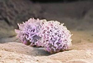
Background imageMacrophage Collection: Macrophage cells, SEM

Macrophage cells, SEM
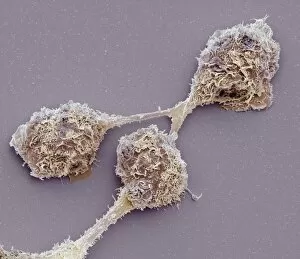
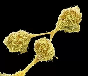
Macrophage cells. Coloured scanning electron micrograph (SEM) of macrophage white blood cells. Macrophages are cells of the bodys immune system

Background imageMacrophage Collection: Bacteria infecting a macrophage, SEM

Bacteria infecting a macrophage, SEM
Bacteria infecting a macrophage. Coloured scanning electron micrograph (SEM) of Mycobacterium tuberculosis bacteria (purple) infecting a macrophage white blood cell

Background imageMacrophage Collection: Macrophage engulfing bead

Macrophage engulfing bead
Illustration based on a scanning electron micrograph of a human macrophage phagocytosing (engulfing) a latex bead. Macrophages are monocytes from circulating blood that have migrated to tissues

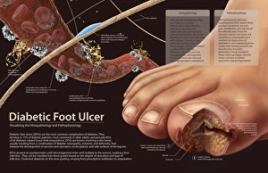
Background imageMacrophage Collection: Histopathology and pathophysiology of diabetic food ulcers

Histopathology and pathophysiology of diabetic food ulcers

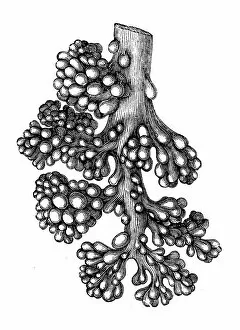
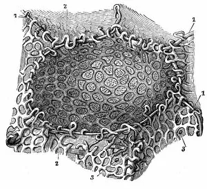
Background imageMacrophage Collection: Lung alveoli tree

Lung alveoli tree
Antique illustration of a Lung alveoli tree

Background imageMacrophage Collection: HIV infected macrophage, SEM C018 / 8598

HIV infected macrophage, SEM C018 / 8598
HIV infected macrophage. Coloured ion-abrasion scanning electron micrograph (IA-SEM) of a macrophage white blood cell infected with human immunodeficiency virus (HIV, red)

Background imageMacrophage Collection: White blood cell antigen presentation C016 / 9058

White blood cell antigen presentation C016 / 9058
White blood cell antigen presentation. Coloured scanning electron micrograph (SEM) showing the interaction between a macrophage (yellow) and a T helper lymphocyte (Th cell, green)

Background imageMacrophage Collection: Macrophage white blood cells, TEM

Macrophage white blood cells, TEM
Macrophage white blood cells. Transmission electron micrograph (TEM) of a section through two macrophages located within connective tissue, showing the nuclei and numerous lysosomes (dark spots)

Background imageMacrophage Collection: Macrophage and lymphocytes, TEM

Macrophage and lymphocytes, TEM
Macrophage and lymphocytes, transmission electron micrograph (TEM). Macrophage surrounded by lymphocytes in a lymph node. This view includes many intercellular contacts

Background imageMacrophage Collection: Macrophages, SEM

Macrophages, SEM
Macrophages. Coloured scanning electron micrograph (SEM) of macrophage white blood cells. Macrophages are cells of the bodys immune system

Background imageMacrophage Collection: Pulmonary alveolus

Pulmonary alveolus
Antique illustration of a Pulmonary alveolus

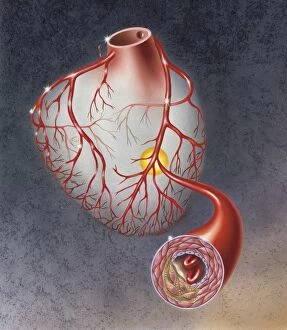
Background imageMacrophage Collection: Heart with arteries showing cholesterol in one and plaque in the other

Heart with arteries showing cholesterol in one and plaque in the other
Heart with arteries showing cholesterol in one artery and atherosclerotic plaque in the other

Background imageMacrophage Collection: Arteries on heart showing atherosclerotic plaque in an artery

Arteries on heart showing atherosclerotic plaque in an artery

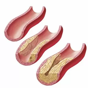
Background imageMacrophage Collection: Build-up of atherosclerotic plaque in an artery

Build-up of atherosclerotic plaque in an artery

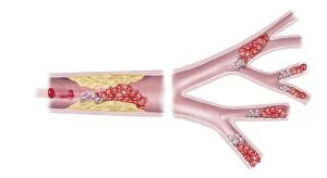
Background imageMacrophage Collection: Acute coronary syndrome - microvascular obstruction

Acute coronary syndrome - microvascular obstruction

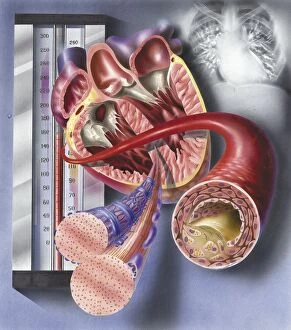
Background imageMacrophage Collection: Interior view of heart with detail of muscle cells and atherosclerotic artery

Interior view of heart with detail of muscle cells and atherosclerotic artery. Chest x-ray in background

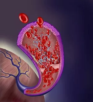
Background imageMacrophage Collection: Thrombus forming on valve within vein

Thrombus forming on valve within vein

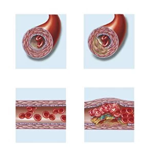
Background imageMacrophage Collection: Normal artery compared to plaque and thrombus formation in artery

Normal artery compared to plaque and thrombus formation in artery

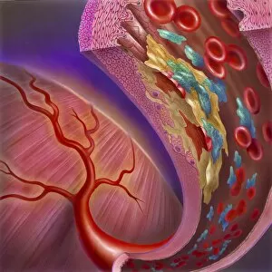
Background imageMacrophage Collection: Artery showing atherosclerotic plaque, platelets and red blood cells

Artery showing atherosclerotic plaque, platelets and red blood cells
Close-up of artery showing atherosclerotic plaque, platelets and red blood cells

Background imageMacrophage Collection: Conceptual image of cancer virus

Conceptual image of cancer virus

Background imageMacrophage Collection: Microscopic view of a group of macrophages

Microscopic view of a group of macrophages. Macrophages contribute to tumor growth and progression. Attracted to oxygen-starved (hypoxic) and necrotic tumor cells they promote chronic inflammation

Background imageMacrophage Collection: 3D rendering of macrophage phagocytosis

3D rendering of macrophage phagocytosis

Background imageMacrophage Collection: Microscopic view of phagocytic macrophages

Microscopic view of phagocytic macrophages, which are involved in the immune response within the body

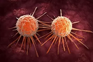
Background imageMacrophage Collection: Microscopic view of cancer virus

Microscopic view of cancer virus

Background imageMacrophage Collection: Isolated cancer cell Macrophage

Isolated cancer cell Macrophage. Macrophages contribute to tumor growth and progression. Attracted to oxygen-starved (hypoxic) and necrotic tumor cells they promote chronic inflammation

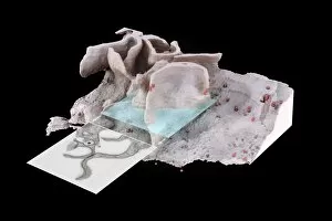
Background imageMacrophage Collection: Surface of HIV infected microphage

Surface of HIV infected microphage
3D representation of the surface and interior of an HIV-infected macrophage obtained using newly developed tools for 3D imaging using ion-abrasion scanning electron microscopy

Background imageMacrophage Collection: Torn muscle fibers with healing stages surrounding

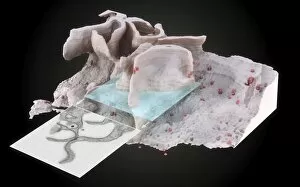
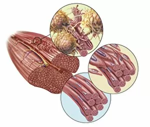
Torn muscle fibers with healing stages surrounding. 1. Macrophage clean-up. 2. New cells migrate to repair. 3. Cells differentiate to become muscle cells

Background imageMacrophage Collection: Human Digestive System, Macrophage under microscope

Human Digestive System, Macrophage under microscope
Medicine - Human Histology - Digestive system - Macrophage under microscope

Background imageMacrophage Collection: Activated macrophage, SEM F006 / 8635

Activated macrophage, SEM F006 / 8635
Activated human macrophage, coloured scanning electron micrograph (SEM). Magnification: x2, 700 when printed at 10 centimetres wide

Background imageMacrophage Collection: Activated macrophage, SEM F006 / 8634

Activated macrophage, SEM F006 / 8634
Activated human macrophage, coloured scanning electron micrograph (SEM). Magnification: x2, 700 when printed at 10 centimetres wide

Background imageMacrophage Collection: Microglial white blood cell, SEM C016 / 9115

Microglial white blood cell, SEM C016 / 9115
Microglial white blood cell. Coloured scanning electron micrograph (SEM) of a microglial cell. Microglia are phagocytic macrophage white blood cells that are the main form of immune system in

Background imageMacrophage Collection: Shigella bacterium and macrophage, SEM C016 / 8922

Shigella bacterium and macrophage, SEM C016 / 8922
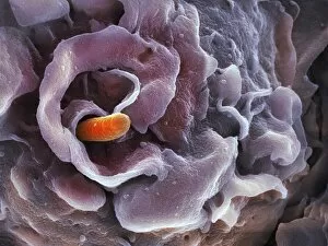
Shigella bacterium. Coloured scanning electron micrograph (SEM) of a Shigella sp. bacterium (orange) being engulfed by a macrophage white blood cell. This process is called phagocytosis

Background imageMacrophage Collection: Shigella bacterium and macrophage, SEM C016 / 8924

Shigella bacterium and macrophage, SEM C016 / 8924
Shigella bacterium. Coloured scanning electron micrograph (SEM) of a Shigella sp. bacterium (green) being engulfed by a macrophage white blood cell. This process is called phagocytosis

Background imageMacrophage Collection: Microglial white blood cell, SEM C016 / 9119

Microglial white blood cell, SEM C016 / 9119
Microglial white blood cell. Coloured scanning electron micrograph (SEM) of a microglial cell. Microglia are phagocytic macrophage white blood cells that are the main form of immune system in

Background imageMacrophage Collection: Microglial white blood cell, SEM C016 / 9117

Microglial white blood cell, SEM C016 / 9117
Microglial white blood cell. Coloured scanning electron micrograph (SEM) of a microglial cell. Microglia are phagocytic macrophage white blood cells that are the main form of immune system in

Background imageMacrophage Collection: Microglial white blood cell, SEM C016 / 9109

Microglial white blood cell, SEM C016 / 9109
Microglial white blood cell. Coloured scanning electron micrograph (SEM) of a microglial cell. Microglia are phagocytic macrophage white blood cells that are the main form of immune system in

Background imageMacrophage Collection: Shigella bacterium and macrophage, SEM C016 / 8923

Shigella bacterium and macrophage, SEM C016 / 8923
Shigella bacterium. Coloured scanning electron micrograph (SEM) of a Shigella sp. bacterium (orange) being engulfed by a macrophage white blood cell. This process is called phagocytosis

Background imageMacrophage Collection: White blood cell antigen presentation C016 / 9057

White blood cell antigen presentation C016 / 9057
White blood cell antigen presentation. Coloured scanning electron micrograph (SEM) showing the interaction between a macrophage (red) and a T helper lymphocyte (Th cell, yellow)

Background imageMacrophage Collection: Microglial white blood cell, SEM C016 / 9114

Microglial white blood cell, SEM C016 / 9114
Microglial white blood cell. Coloured scanning electron micrograph (SEM) of a microglial cell. Microglia are phagocytic macrophage white blood cells that are the main form of immune system in

Background imageMacrophage Collection: Microglial white blood cells, SEM C016 / 9113

Microglial white blood cells, SEM C016 / 9113
Microglial white blood cells. Coloured scanning electron micrograph (SEM) of microglial cells. Microglia are phagocytic macrophage white blood cells that are the main form of immune system in

Background imageMacrophage Collection: Microglial white blood cell, SEM C016 / 9118

Microglial white blood cell, SEM C016 / 9118
Microglial white blood cell. Coloured scanning electron micrograph (SEM) of a microglial cell. Microglia are phagocytic macrophage white blood cells that are the main form of immune system in

Background imageMacrophage Collection: Microglial white blood cells, SEM C016 / 9111

Microglial white blood cells, SEM C016 / 9111
Microglial white blood cells. Coloured scanning electron micrograph (SEM) of microglial cells. Microglia are phagocytic macrophage white blood cells that are the main form of immune system in

Background imageMacrophage Collection: Microglial white blood cells, SEM C016 / 9112

Microglial white blood cells, SEM C016 / 9112
Microglial white blood cells. Coloured scanning electron micrograph (SEM) of microglial cells. Microglia are phagocytic macrophage white blood cells that are the main form of immune system in

Background imageMacrophage Collection: Microglial white blood cell, SEM C016 / 9116

Microglial white blood cell, SEM C016 / 9116
Microglial white blood cell. Coloured scanning electron micrograph (SEM) of a microglial cell. Microglia are phagocytic macrophage white blood cells that are the main form of immune system in

Background imageMacrophage Collection: Lung silicosis, light micrograph C015 / 7129

Lung silicosis, light micrograph C015 / 7129
Lung silicosis. Light micrograph of a section through a lung with silicosis, a type of pneumoconiosis caused by exposure to silica dusts. The lung tissue has become fibrous (centre)

Background imageMacrophage Collection: Microglial white blood cell, SEM C019 / 0247

Microglial white blood cell, SEM C019 / 0247
Microglial white blood cell, SEM

Background imageMacrophage Collection: Microglial white blood cell, SEM C019 / 0246

Microglial white blood cell, SEM C019 / 0246
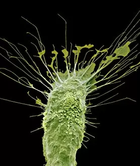
Microglia. Coloured scanning electron micrograph (SEM) of an activated microglial white blood cell. This immune system cell is found in the brain and spinal cord

Background imageMacrophage Collection: Microglial white blood cell, SEM C019 / 0245

Microglial white blood cell, SEM C019 / 0245
Microglia. Coloured scanning electron micrograph (SEM) of an activated microglial white blood cell. This immune system cell is found in the brain and spinal cord

Background imageMacrophage Collection: Activated macrophage, SEM C015 / 6377

Activated macrophage, SEM C015 / 6377
Activated macrophage. Coloured scanning electron micrograph (SEM) of an active macrophage white blood cell. Macrophages are cells of the bodys immune system that are found in the tissues rather than

Background imageMacrophage Collection: Activated macrophage, SEM C015 / 6376

Activated macrophage, SEM C015 / 6376
Activated macrophage. Coloured scanning electron micrograph (SEM) of an active macrophage white blood cell. Macrophages are cells of the bodys immune system that are found in the tissues rather than

Background imageMacrophage Collection: Macrophage engulfing TB bacteria, SEM C014 / 0596

Macrophage engulfing TB bacteria, SEM C014 / 0596
Macrophage engulfing TB bacteria. Coloured scanning electron micrograph (SEM) of a macrophage white blood cell engulfing a tuberculosis (Mycobacterium tuberculosis) bacterium (orange)

Background imageMacrophage Collection: Macrophage engulfing TB bacteria, SEM C014 / 0598

Macrophage engulfing TB bacteria, SEM C014 / 0598
Macrophage engulfing TB bacteria. Coloured scanning electron micrograph (SEM) of a macrophage white blood cell engulfing a tuberculosis (Mycobacterium tuberculosis) bacterium (purple)

Background imageMacrophage Collection: Macrophage engulfing TB bacteria, SEM C014 / 0595

Macrophage engulfing TB bacteria, SEM C014 / 0595
Macrophage engulfing TB bacteria. Coloured scanning electron micrograph (SEM) of a macrophage white blood cell engulfing a tuberculosis (Mycobacterium tuberculosis) bacterium (pink)

Background imageMacrophage Collection: Macrophage engulfing TB bacteria, SEM C014 / 0594

Macrophage engulfing TB bacteria, SEM C014 / 0594
Macrophage engulfing TB bacteria. Coloured scanning electron micrograph (SEM) of a macrophage white blood cell engulfing a tuberculosis (Mycobacterium tuberculosis) bacterium (yellow)

Background imageMacrophage Collection: Macrophage engulfing TB bacteria, SEM C014 / 0593

Macrophage engulfing TB bacteria, SEM C014 / 0593
Macrophage engulfing TB bacteria. Coloured scanning electron micrograph (SEM) of a macrophage white blood cell engulfing a tuberculosis (Mycobacterium tuberculosis) bacterium (purple)

Background imageMacrophage Collection: Macrophage engulfing TB bacteria, SEM C014 / 0592

Macrophage engulfing TB bacteria, SEM C014 / 0592
Macrophage engulfing TB bacteria. Coloured scanning electron micrograph (SEM) of a macrophage white blood cell engulfing a tuberculosis (Mycobacterium tuberculosis) bacterium (orange)

Background imageMacrophage Collection: Macrophage engulfing TB bacteria, SEM C014 / 0590

Macrophage engulfing TB bacteria, SEM C014 / 0590
Macrophage engulfing TB bacteria. Coloured scanning electron micrograph (SEM) of a macrophage white blood cell engulfing a tuberculosis (Mycobacterium tuberculosis) bacterium (blue)

Background imageMacrophage Collection: Macrophage engulfing TB bacteria, SEM C014 / 0589

Macrophage engulfing TB bacteria, SEM C014 / 0589
Macrophage engulfing TB bacteria. Coloured scanning electron micrograph (SEM) of a macrophage white blood cell engulfing a tuberculosis (Mycobacterium tuberculosis) bacterium (yellow)

Background imageMacrophage Collection: Macrophage engulfing TB bacteria, SEM C014 / 0591

Macrophage engulfing TB bacteria, SEM C014 / 0591
Macrophage engulfing TB bacteria. Coloured scanning electron micrograph (SEM) of a macrophage white blood cell engulfing a tuberculosis (Mycobacterium tuberculosis) bacterium (bright green)

Background imageMacrophage Collection: Macrophage engulfing TB bacteria, SEM C017 / 8309

Macrophage engulfing TB bacteria, SEM C017 / 8309
Macrophage engulfing TB bacteria. Coloured scanning electron micrograph (SEM) of a macrophage white blood cell engulfing tuberculosis (Mycobacterium tuberculosis) bacteria (pink)

Background imageMacrophage Collection: Macrophage feeding bacteria, artwork C017 / 8247

Macrophage feeding bacteria, artwork C017 / 8247
Computer artwork of a macrophage (blue), ingesting a bacterium (red), a process known as phagocytosis. The nucleus is depicted in green

Background imageMacrophage Collection: Lymph node, light micrograph C015 / 6223

Lymph node, light micrograph C015 / 6223
Lymph node. Light micrograph of a section through a lymph node. The blue tissue at right is the fibrous outer capsule of the node. Beneath it is the subcapsular sinus (white)

Background imageMacrophage Collection: Lymph node, light micrograph C015 / 6222

Lymph node, light micrograph C015 / 6222
Lymph node. Light micrograph of a section through a lymph node. The blue tissue at left is the fibrous outer capsule of the node. Beneath it is the subcapsular sinus (white)

Background imageMacrophage Collection: Antigen presentation, SEM C016 / 3105

Antigen presentation, SEM C016 / 3105
Antigen presentation. Coloured scanning electron micrograph (SEM) showing the interaction between a macrophage (blue) and a T helper lymphocyte (Th cell, yellow)

Background imageMacrophage Collection: Antigen presentation, SEM C016 / 3104

Antigen presentation, SEM C016 / 3104
Antigen presentation. Coloured scanning electron micrograph (SEM) showing the interaction between a macrophage (brown) and a T helper lymphocyte (Th cell, red)

Background imageMacrophage Collection: Macrophage and platelets, SEM C016 / 3094

Macrophage and platelets, SEM C016 / 3094
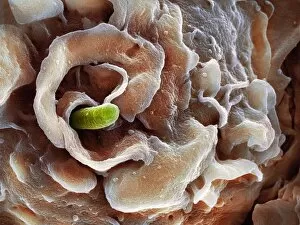
Macrophage and platelets. Coloured scanning electron micrograph (SEM) of platelets (green) attached to a macrophage white blood cell (turquoise)

Background imageMacrophage Collection: E. coli induced cell death, SEM C016 / 3078

E. coli induced cell death, SEM C016 / 3078
E. coli induced cell death. Coloured scanning electron micrograph (SEM) of a macrophage white blood cell (centre) that is being destroyed by toxins released by Escherichia coli (E)

Background imageMacrophage Collection: E. coli induced cell death, SEM C016 / 3077

E. coli induced cell death, SEM C016 / 3077
E. coli induced cell death. Coloured scanning electron micrograph (SEM) of a macrophage white blood cell (centre) that is being destroyed by toxins released by Escherichia coli (E)

Background imageMacrophage Collection: Macrophage engulfing a nerve cell, TEM C013 / 4803

Macrophage engulfing a nerve cell, TEM C013 / 4803
Macrophage engulfing a nerve cell. Transmission electron micrograph (TEM) of a section through a macrophage white blood cell (blue) that has engulfed a nerve cell (neuron, centre)

Background imageMacrophage Collection: Coloured SEM of liver cells and bile canaliculi

Coloured SEM of liver cells and bile canaliculi

Background imageMacrophage Collection: F. colour SEM of macrophage & blood cells in liver

F. colour SEM of macrophage & blood cells in liver
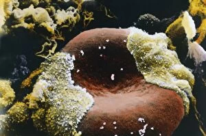
Liver. False-colour scanning electron micrograph of a section of liver tissue showing a group of red blood cells and a macrophage (yellow) in a capillary

Background imageMacrophage Collection: Coloured SEM of blood vessels in the skin

Coloured SEM of blood vessels in the skin
Skin blood vessels. Coloured scanning electron micrograph (SEM) of two small blood vessels (far left and upper right) in the dermis of the skin

Background imageMacrophage Collection: Dendritic cell, computer artwork

Dendritic cell, computer artwork
Dendritic cells. Computer artwork of dendritic cells, part of the human immune system. These are a type of macrophage cell found in the bodys tissues

Background imageMacrophage Collection: Dendritic cell, TEM

Dendritic cell, TEM
Dendritic cell. Coloured transmission electron micrograph (TEM) of a section through dendritic cells. These are part of the human immune system

Background imageMacrophage Collection: White blood cell types

White blood cell types
Artwork featuring different types of white blood cells that comprise the orchestra of the human immune system, the bodys defence mechanism

Background imageMacrophage Collection: Immune system components

Immune system components
Immune system. Conceptual computer artwork of a man standing with his arms outstretched surrounded by components of the immune system. The skeletal system is seen, with the joints highlighted in red

Background imageMacrophage Collection: Lymph node, SEM

Lymph node, SEM
Lymph node. Coloured scanning electron micrograph (SEM) of a freeze fracture section through a lymph node, showing part of the medullary sinus

Background imageMacrophage Collection: Lymph node

Lymph node
Illustration of a sectioned, whole human lymph node. Lymph nodes are encapsulated, kidney shaped structures that are usually located at intervals along lymph vessels

Background imageMacrophage Collection: Macrophage engulfing bacteria, artwork

Macrophage engulfing bacteria, artwork
Macrophage engulfing bacteria. Computer artwork of a macrophage white blood cell (pink) engulfing bacteria (blue). This process is called phagocytosis


All Professionally Made to Order for Quick Shipping

Macrophages, the mighty defenders of our immune system, play a crucial role in protecting us from harmful invaders. These incredible cells are like superheroes, constantly on guard to keep us healthy. In stunning artwork and microscopic images captured by powerful electron microscopes, we get a glimpse into the fascinating world of macrophages. Dendritic cells, another type of immune cell closely related to macrophages, are depicted in beautiful artwork that showcases their intricate structure. A transmission electron microscope (TEM) image reveals the detailed morphology of a human macrophage. Its complex network of cytoplasmic extensions is truly mesmerizing. In another scanning electron microscope (SEM) image, we witness bacteria infecting a macrophage - an intense battle between pathogen and defender. The SEM also captures the moment when a tuberculosis bacterium is engulfed by a determined macrophage. This remarkable act demonstrates how these cells tirelessly eliminate threats within our bodies. An activated macrophage is showcased in yet another SEM image - its surface covered with tiny protrusions called filopodia as it prepares for action against invading pathogens or damaged tissue. But it's not just infectious agents that fall victim to the might of these phagocytic warriors; even non-living objects like beads can be engulfed by them. The versatility and efficiency of macrophages never cease to amaze. Unfortunately, sometimes our own immune system can turn against itself. In multiple sclerosis patients, SEM images reveal abnormalities in myelin-producing cells caused by misguided attacks from malfunctioning immune cells such as macrophages. As we delve deeper into understanding these remarkable guardians of health through TEM images showcasing their cellular structures and functions, we gain valuable insights into how they work tirelessly day and night to keep us safe from harm. From battling bacterial infections to defending against autoimmune diseases like multiple sclerosis – this captivating journey through various microscopy techniques highlights the extraordinary capabilities possessed by these essential components of our immune system.

© Copyright Media Storehouse, All Rights Reserved 2003 - 2024
Facebook     Pinterest     100% Payment Secure     Reviews IO