
Macula Collection
The macula, a tiny yet crucial part of the human eye, holds immense significance in our visual perception


All Professionally Made to Order for Quick Shipping
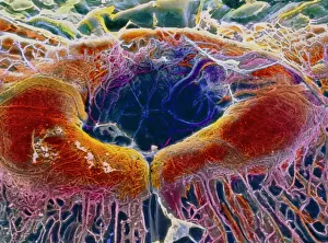
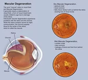
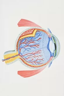
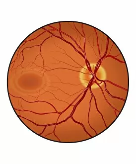
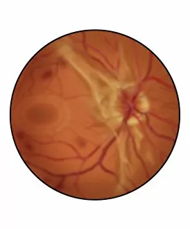
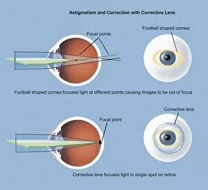
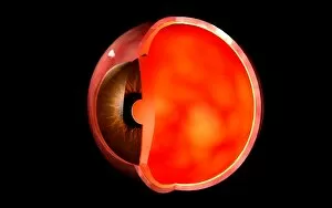
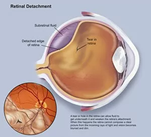
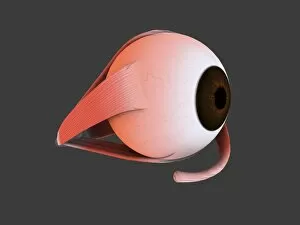
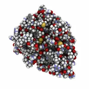
The macula, a tiny yet crucial part of the human eye, holds immense significance in our visual perception. This false-colour scanning electron microscope (SEM) image of the retina featuring the central fovea showcases the intricate beauty and complexity of this remarkable structure. In this captivating cross-section diagram of the human eye, we can observe how the macula is positioned within its anatomical context. Its location at the center of our vision allows for sharp focus and detailed clarity when viewing objects directly in front of us. This biomedical illustration provides an insightful glimpse into the anatomy of our eyes, with particular emphasis on the macula. The conceptual image beautifully depicts how this small area plays a vital role in capturing light and transmitting it to our brain for interpretation. Astonishingly, even though minuscule in size, any impairment or degeneration within the they are have profound consequences on our sight. This retina with macular degeneration serves as a stark reminder that protecting and caring for this delicate region is essential for maintaining optimal vision throughout life. As we delve deeper into understanding human eye anatomy through conceptual images like these, we gain appreciation for how intricately nature has designed such a complex organ. The interplay between different structures becomes evident as we explore this conceptual image depicting both the eye and skull together. An orbital cut revealing not only nerves but also highlighting key components such as ciliary ganglion and oculomotor nerve further emphasizes just how interconnected each element is within our visual system. It reminds us that proper functioning of every part contributes to seamless vision. Through these fascinating visuals showcasing various aspects related to macula's position within human eye anatomy – from cross-sections to conceptual illustrations – we are reminded once again about its importance in shaping our ability to perceive and interpret what lies before us visually. Understanding more about "macula" helps us appreciate both its fragility and resilience while urging us to prioritize regular check-ups and care to ensure its optimal health.