Fraud Blocker Skip to main content

Magnification Collection (#23)

Magnification: Revealing the Hidden World From the intricate patterns of a snowflake to the delicate structure of a human cell

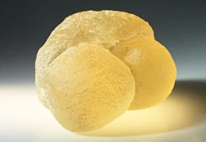
Background imageMagnification Collection: Model of pine pollen

Model of pine pollen

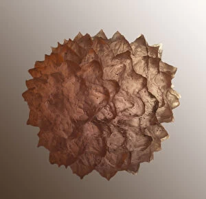
Background imageMagnification Collection: Model of ragweed pollen

Model of ragweed pollen

Background imageMagnification Collection: Magnification of grain of schist rock

Magnification of grain of schist rock

Background imageMagnification Collection: Magnification of grain of gneiss rock

Magnification of grain of gneiss rock

Background imageMagnification Collection: Magnification of grain of sandstone rock

Magnification of grain of sandstone rock

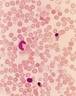
Background imageMagnification Collection: Medicine - anatomy - cardio-circulatory system - blood smear with sickle-shaped red blood cells

Medicine - anatomy - cardio-circulatory system - blood smear with sickle-shaped red blood cells
Medicine - Anatomy - Cardio-circulatory system - Blood smear with sickle-shaped red blood cells, typical of Mediterranean anemia or Cooleys disease, at the microscope

Background imageMagnification Collection: Medicine - anatomy - cardio-circulatory system - human blood smear, microscope view

Medicine - anatomy - cardio-circulatory system - human blood smear, microscope view
Medicine - Anatomy - Cardio-circulatory system - Human blood smear

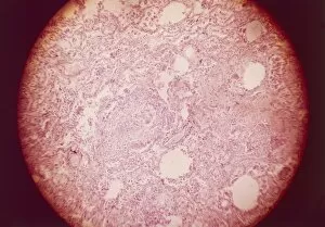
Background imageMagnification Collection: Chronic hepatitis

Chronic hepatitis
Medicine - Pathology - Chronic hepatitis

Background imageMagnification Collection: Respiratory system, Lung, Tuberculosis, Pulmonary phthisis

Respiratory system, Lung, Tuberculosis, Pulmonary phthisis
Medicine - Anatomy - Pathology - Respiratory system - Lung - Tuberculosis - Pulmonary phthisis

Background imageMagnification Collection: Histological specimen of a liver of a cardiopath in which the liver parenchyma is atrophic due to

Histological specimen of a liver of a cardiopath in which the liver parenchyma is atrophic due to blood stasis. X150
Medicine. Histological specimen of a liver of a cardiopath in which the liver parenchyma is atrophic due to blood stasis. x150

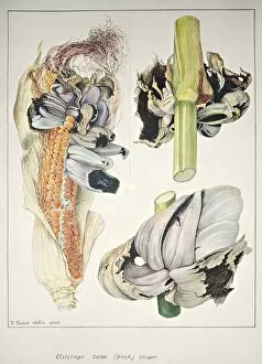
Background imageMagnification Collection: Botany, Plant pathology, Tumors of maize or corn smut, caused by fungus Ustilago maydis

Botany, Plant pathology, Tumors of maize or corn smut, caused by fungus Ustilago maydis
Botany - Plant pathology - Tumors of maize or corn smut, caused by fungus Ustilago maydis

Background imageMagnification Collection: Model of a bacterium, close-up

Model of a bacterium, close-up

Background imageMagnification Collection: Model of vorticella

Model of vorticella

Background imageMagnification Collection: Botany, Fungi, Mycorrhizal fungus, hypha in cell, illustration

Botany, Fungi, Mycorrhizal fungus, hypha in cell, illustration
Botany - Fungi - Mycorrhizal fungus, hypha in cell, illustration

Background imageMagnification Collection: Botany, Algae, Bacillariophyceae, Diatom reproduction, illustration

Botany, Algae, Bacillariophyceae, Diatom reproduction, illustration
Botany - Algae - Bacillariophyceae - Diatom reproduction, illustration

Background imageMagnification Collection: Botany, Apiaceae, Section of fruits of Coriander Coriandrum sativum; Hemlock Conium maculatum;

Botany, Apiaceae, Section of fruits of Coriander Coriandrum sativum; Hemlock Conium maculatum; Parsley Petroselinum
Botany - Apiaceae - Section of fruits of Coriander (Coriandrum sativum); Hemlock (Conium maculatum); Parsley (Petroselinum sativum)

Background imageMagnification Collection: Botany, Fungi, Phycomycetes, fertilization of Saprolegnia

Botany, Fungi, Phycomycetes, fertilization of Saprolegnia
Botany - Fungi - Phycomycetes, fertilization of Saprolegnia

Background imageMagnification Collection: Biology, Various forms of Chloroplasts

Biology, Various forms of Chloroplasts
Biology - Various forms of Chloroplasts

Background imageMagnification Collection: Biology, Plant cell structure, cross section, Illustration

Biology, Plant cell structure, cross section, Illustration
Biology - Plant cell structure. Illustration, section

Background imageMagnification Collection: Botany, Dicotyledons, Upper part of style and stigma, illustration

Botany, Dicotyledons, Upper part of style and stigma, illustration
Botany - Dicotyledons - Upper part of style and stigma. Illustration

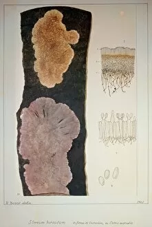
Background imageMagnification Collection: Botany, Plant pathology, Stereum hirsutum on hackberrys bark, illustration

Botany, Plant pathology, Stereum hirsutum on hackberrys bark, illustration
Botany - Plant pathology - Stereum hirsutum on hackberrys bark. Illustration

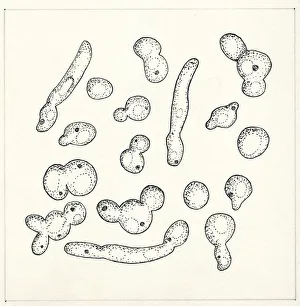
Background imageMagnification Collection: Botany, Fungi, Yeast Candida tropicalis, illustration

Botany, Fungi, Yeast Candida tropicalis, illustration
Botany - Fungi - Yeast (Candida tropicalis), illustration

Background imageMagnification Collection: Botany, Fungi, Acrasiomycetes, Cell of Labyrinthula Macrocystis, illustration

Botany, Fungi, Acrasiomycetes, Cell of Labyrinthula Macrocystis, illustration
Botany - Fungi - Acrasiomycetes - Cell of Labyrinthula Macrocystis

Background imageMagnification Collection: Botany, Biology, Plant histology, Fungi, Ascomycota, Penicillium crustaceum, cleistothecia

Botany, Biology, Plant histology, Fungi, Ascomycota, Penicillium crustaceum, cleistothecia, illustration
Botany - Biology - Plant histology - Fungi - Ascomycota - Penicillium crustaceum, cleistothecia. Illustration

Background imageMagnification Collection: Botany, Fungi, Ascomycota, Penicilliums, soil fungi, illustration

Botany, Fungi, Ascomycota, Penicilliums, soil fungi, illustration
Botany - Fungi - Ascomycota - Penicilliums, soil fungi. Illustration

Background imageMagnification Collection: Biology, Leaf of Dicotyledons, cross section, illustration

Biology, Leaf of Dicotyledons, cross section, illustration
Biology - Leaf of Dicotyledons. Illustration, cross section

Background imageMagnification Collection: Ruddy Darter (Sympetrum sanguineum), magnification of male dragonfly head

Ruddy Darter (Sympetrum sanguineum), magnification of male dragonfly head

Background imageMagnification Collection: Using USB microscope to display magnified image of caterpillar on laptop screen

Using USB microscope to display magnified image of caterpillar on laptop screen

Background imageMagnification Collection: Queen wasp on a dead thistle flower head

Queen wasp on a dead thistle flower head

Background imageMagnification Collection: Model of a bee, overhead view

Model of a bee, overhead view

Background imageMagnification Collection: Ichneumon wasp, side view

Ichneumon wasp, side view

Background imageMagnification Collection: Model of cyclops

Model of cyclops

Background imageMagnification Collection: Honey bee (Apis mellifera), overhead view

Honey bee (Apis mellifera), overhead view

Background imageMagnification Collection: Model of Tardigrade, side view

Model of Tardigrade, side view

Background imageMagnification Collection: Magnified view of a wasps head

Magnified view of a wasps head

Background imageMagnification Collection: A fly, feeding, side view

A fly, feeding, side view

Background imageMagnification Collection: Magnification of butterfly showing eyes, proboscis, antennae, and legs

Magnification of butterfly showing eyes, proboscis, antennae, and legs

Background imageMagnification Collection: Flea, close-up, side view

Flea, close-up, side view

Background imageMagnification Collection: Flea on blade of grass, close-up

Flea on blade of grass, close-up

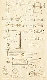
Background imageMagnification Collection: Reflaction and Telescope Optics C017 / 3507

Reflaction and Telescope Optics C017 / 3507
Plate from 18th century encyclopedia showing diagrams of telescope optics, and light raypaths through prisms and lenses to the eye

Background imageMagnification Collection: Diamond refracting light F008 / 3208

Diamond refracting light F008 / 3208
Diamond refracting light

Background imageMagnification Collection: Reading glasses F008 / 2082

Reading glasses F008 / 2082
Reading glasses

Background imageMagnification Collection: Skeletal muscle fibres, light micrograph

Skeletal muscle fibres, light micrograph
Skeletal muscle fibres. Light micrograph of skeletal muscle fibres showing the characteristic banding. Cell nuclei are yellow and red blood cells, in capillaries in the endomycium, are red

Background imageMagnification Collection: Rosuvastatin cholesterol-lowering drug

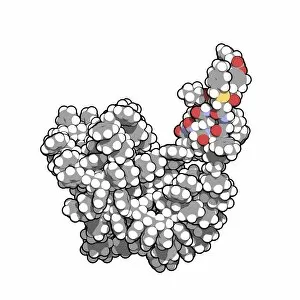
Rosuvastatin cholesterol-lowering drug, molecular model. This is statin class works by blocking the effects of the enzyme HMG-CoA reductase

Background imageMagnification Collection: Insulin A chain molecule

Insulin A chain molecule
Insulin A chain. Computer model of an A chain of human insulin that has been synthesized on a crosslinked polystyrene solid support. This is an example of solid phase peptide synthesis (SPPS)

Background imageMagnification Collection: Coccidiosis, light micrograph F005 / 6082

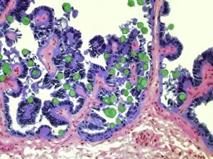
Coccidiosis, light micrograph F005 / 6082
Coccidiosis. Light micrograph of a section through an hepatic bile duct infected with cocidian protozoa (green). Magnification: x300 when printed at 10 centimetres wide

Background imageMagnification Collection: Transfer RNA-synthetase complex molecule

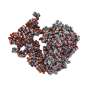
Transfer RNA-synthetase complex molecule. Molecular model of a human tryptophanyl-tRNA synthetase molecule (red) complexed with a tRNA(Trp) molecule (blue)

Background imageMagnification Collection: Roundworm ovaries, light micrograph

Roundworm ovaries, light micrograph
Roundworm ovaries. Light micrograph of a section through ovaries from an Ascaris sp. roundworm. Magnification: x100 when printed at 10 centimetres wide


All Professionally Made to Order for Quick Shipping

Magnification: Revealing the Hidden World From the intricate patterns of a snowflake to the delicate structure of a human cell, magnification allows us to explore and unravel the mysteries of our world. Peering through the lens, we uncover secrets that would otherwise remain unseen. Take for instance Crysotile asbestos, its fibrous nature exposed under intense magnification, reminding us of its hidden dangers lurking within. In medical illustrations, an appendix with appendicitis comes alive as we zoom in closer. The inflamed tissues become vividly apparent, highlighting the urgency for surgical intervention. Moving deeper into our bodies, we discover the liver - a complex organ responsible for countless vital functions. Under magnification, its intricate network of cells and blood vessels reveals itself like an elaborate cityscape bustling with activity. But it's not just within ourselves that magnification unveils wonders; it extends beyond to nature's realm. A dandelion's fruiting head transforms into a mesmerizing display when observed up close – each tiny seed ready to embark on its own journey through wind and time. Fractals captivate our imagination as well – from Julia fractals with their infinite complexity to Mandelbrot fractals' self-repeating patterns. These mathematical marvels remind us of nature's innate ability to create beauty even in abstract forms. Anton van Leeuwenhoek revolutionized science by observing animalcules through his microscope in 1795. His discoveries opened up new frontiers in microbiology and forever changed our understanding of life at microscopic levels. Even seemingly ordinary creatures like snails hold surprises when examined closely; their teeth reveal intricate structures designed for efficient feeding and survival. Ponds teem with life too - water fleas gracefully glide alongside green algae like Volvox aureus. Magnified images transport us into this miniature ecosystem where every organism plays a crucial role in maintaining balance and harmony. And finally, we come full circle with snowflakes.