
Medical Exam Collection
"Exploring the Depths of Medical Knowledge: A Journey through Time" Under the arches of medical history, we uncover a treasure trove of knowledge


All Professionally Made to Order for Quick Shipping
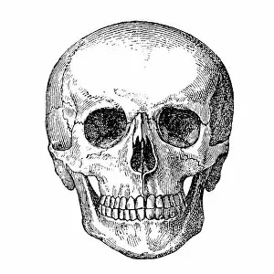
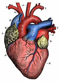
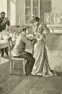
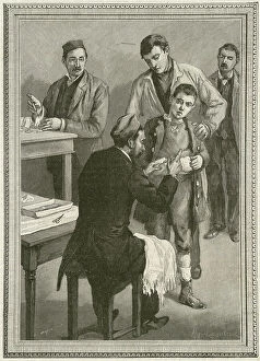
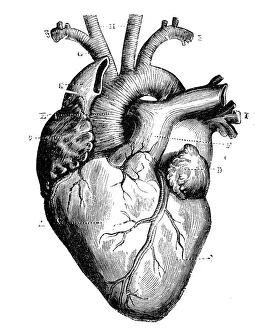
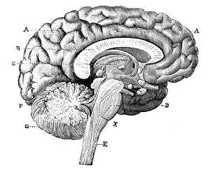
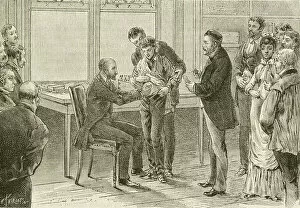
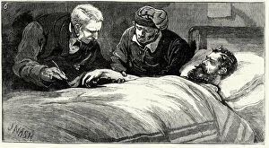
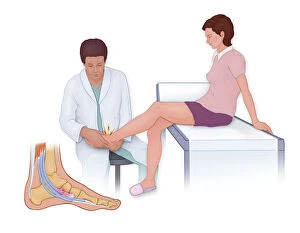
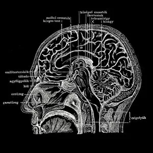
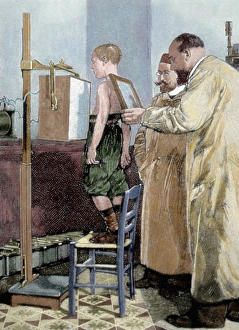
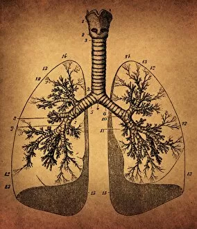
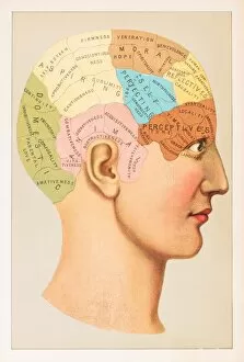
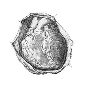
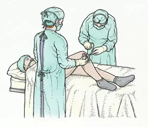
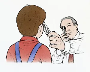
"Exploring the Depths of Medical Knowledge: A Journey through Time" Under the arches of medical history, we uncover a treasure trove of knowledge. From an intriguing brain anatomy engraving dating back to 1895, to a captivating black and white illustration capturing the electrical activity during a petit mal seizure, our understanding of the human body continues to evolve. Delving deeper into this antique scientific world, we encounter high-resolution depictions that mesmerize us. The intricate details of both the heart and brain are unveiled before our eyes, showcasing their complexity and beauty. Intriguingly, it was Wilhelm Conrad Rontgen who revolutionized medicine with his discovery of X-rays in 1895. These rays allowed doctors to peer inside the human body like never before, revealing hidden secrets within. As we journey further into medical examinations throughout time, we witness Doctor Pasteur diligently examining a young boy infected with rabies in the 19th century. This poignant engraving reminds us of the tireless efforts made by physicians to combat diseases that once plagued humanity. Even more intimate exams are captured in cross-section anatomy illustrations – including a rectal exam on a normal male – highlighting how thorough medical assessments have become over time. Finally, our exploration concludes with Ned Kelly's presence at Melbourne's Gaol Hospital. His image serves as a reminder that even notorious figures sought medical care when needed; no one is exempt from health concerns. Through these captivating visuals spanning centuries past, we gain insight into how far medicine has come. Each image tells its own story and invites us to appreciate both the artistry and advancements that shape modern healthcare practices today.