
Medical Imaging Collection
"Unveiling the Invisible: A Journey through Medical Imaging" Step back in time to Ladysmith Hospital Corps


All Professionally Made to Order for Quick Shipping
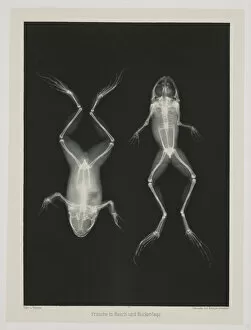
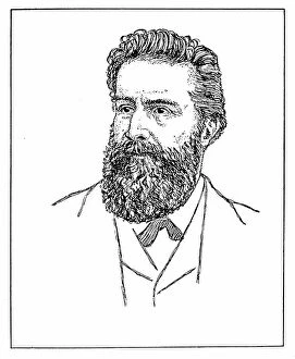
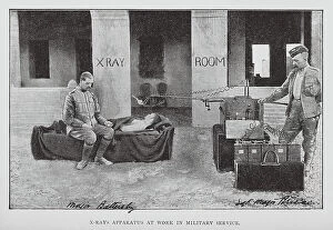
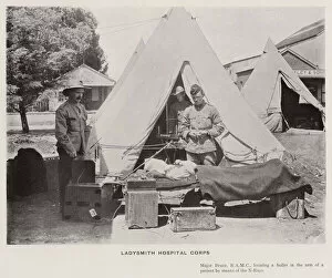
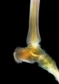
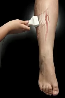
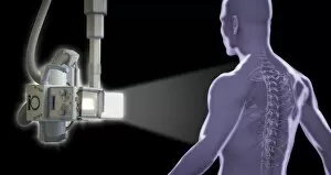
"Unveiling the Invisible: A Journey through Medical Imaging" Step back in time to Ladysmith Hospital Corps, where black and white photos capture the essence of early medical imaging. Witness the birth of Diagnostic Radiology as X-rays reveal two frogs in 1896, marking a revolutionary breakthrough. The application of X-rays knows no bounds - from verifying packages to exploring Ether's discovery by German physicist Wilhelm Conrad Roentgen. With caution, radiologists delve into the realm of X-rays, protecting themselves while peering into the human body's mysteries. Transporting us further into history, an anonymous illustration from 1925 showcases how X-ray technology evolved. It unveils its power to see through objects and bodies alike – a glimpse into an unseen world that captivates even today. Fast forward to modern times; witness the miracle of life unfold before your eyes. A pregnant woman gazes at her baby scan with awe and wonder (F008/2985). The magic continues as another expectant mother cherishes this precious moment (F008/2983). In this captivating journey, we encounter a woman immersed in emotions as she looks at her baby scan (F008/3589). A nurse stands beside her patient with compassion and care (F008/2988), while a couple shares their joyous anticipation together (F008/3586). Medical imaging has come far since its humble beginnings. From Ladysmith Hospital Corps to cutting-edge technologies today, it remains an invaluable tool for doctors worldwide. Through these images captured throughout history, we celebrate how medical imaging continues to shape our understanding of health and humanity itself.