
Metastatic Collection
"Metastatic: The Silent Spread of Cancer" Ovarian cancer, a formidable adversary that often goes undetected until it reaches its advanced stages


All Professionally Made to Order for Quick Shipping
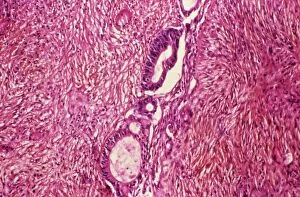
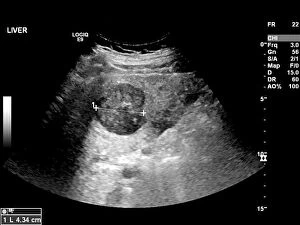
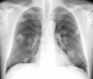
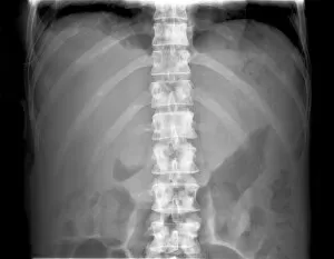
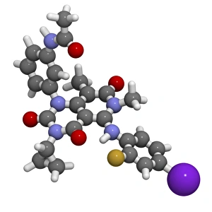
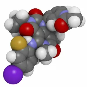
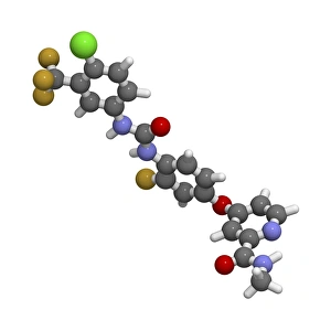
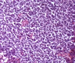
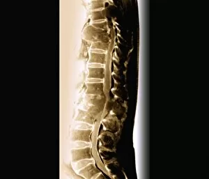
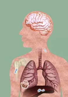
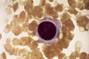
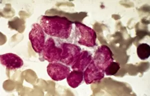
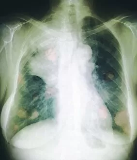
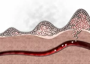
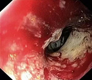
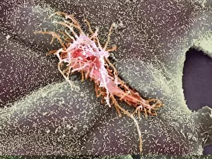
"Metastatic: The Silent Spread of Cancer" Ovarian cancer, a formidable adversary that often goes undetected until it reaches its advanced stages. This light micrograph C015 / 7103 reveals the intricate web cells invading nearby tissues and organs. Secondary lung cancers, stealthily infiltrating the delicate respiratory system. An X-ray unveils their presence, reminding us of the urgent need for early detection and effective treatment. The liver, a vital organ under siege by secondary liver cancer. An ultrasound scan C017 / 7764 uncovers these unwelcome guests, emphasizing the importance of monitoring and managing this aggressive disease. Bones weakened by secondary bone cancer as depicted in an X-ray C017 / 7162. These skeletal invaders cause pain and fractures, highlighting the urgency to address this debilitating condition promptly. A mesmerizing artwork C016 / 9843 portrays a single cancer cell - a reminder that behind every diagnosis lies countless battles fought within our bodies against this relentless disease. Vemurafenib melanoma drug F007 / 0204 offers hope to those battling malignant skin tumors. Its targeted therapy aims to halt the progression of melanoma cells, providing patients with renewed optimism for their future. Trametinib melanoma cancer drug F007 / 0198 emerges as another weapon in our arsenal against aggressive skin cancers. By inhibiting specific signaling pathways within tumor cells, it seeks to disrupt their growth and spread. Regorafenib colorectal cancer drug F007 / 0187 stands at the forefront of innovative treatments for this challenging malignancy. With its ability to target multiple pathways involved in tumor growth, it holds promise for patients seeking new options beyond conventional therapies. Bone cancer's microscopic landscape comes alive through light micrograph F006 / 9810 - a visual representation of how this disease ravages our skeletal structure from within. Early detection remains crucial in combating its devastating effects.