Fraud Blocker Skip to main content

Microbe Collection (page 40)

"Unveiling the Invisible: Exploring the Microbial World" In a realm unseen by the naked eye, lies a fascinating universe teeming with life

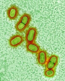
Background imageMicrobe Collection: Legionella bacteria

Legionella bacteria. Coloured transmission electron micrograph (TEM) of four Legionella pneumophila bacteria. The bacteria (pale green) cause Legionnaires disease

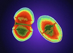
Background imageMicrobe Collection: Staphylococcus epidermidis bacterium

Staphylococcus epidermidis bacterium
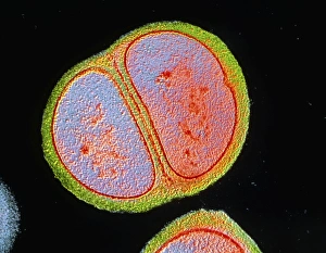
False-colour transmission electron micrograph (TEM) of a single bacterium of Staphylococcus epidermidis undergoing division. Magnification : x17, 500 at 35mm, x38, 500 at 6x7cm size

Background imageMicrobe Collection: Antibiotic action on Staphylococcus

Antibiotic action on Staphylococcus
False-colour transmission electron micrograph (TEM) showing the effect of an antibiotic on the bacterium Staphylococcus aureus

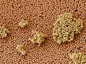
Background imageMicrobe Collection: MRSA bacteria, SEM

MRSA bacteria, SEM
MRSA bacteria, coloured scanning electron micrograph (SEM). Methicillin-resistant Staphylococcus aureus (MRSA) is a Gram-positive, round (coccus) bacterium

Background imageMicrobe Collection: TEM of dividing staphylococcus aureus

TEM of dividing staphylococcus aureus
False-colour transmission electron micrograph (TEM) of Staphylococcus aureus, showing a bacterium in the process of division by binary fission

Background imageMicrobe Collection: MRSA

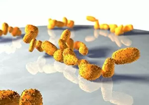
MRSA. Graphic displaying MRSA surrounded by shapes representing bacteria. MRSA stands for methicillin-resistant Staphylococcus aureus. MRSA is a gram-positive, round (coccus) bacterium

Background imageMicrobe Collection: TEM Staphylococcus aureus

TEM Staphylococcus aureus
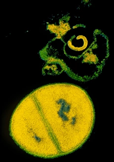
False-colour transmission electron micrograph (TEM) of bacteria Staphylococcus aureus, showing a single bacterium (lower centre)

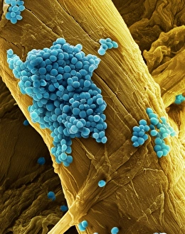
Background imageMicrobe Collection: Staphylococcus aureus bacteria, SEM

Staphylococcus aureus bacteria, SEM
Staphylococcus aureus bacteria, coloured scanning electron micrograph (SEM). These Gram-positive cocci (round) are found on the skin and mucous membranes of humans and many animals

Background imageMicrobe Collection: Yeast cells

Yeast cells, computer artwork. Yeasts are single- cell fungi. Some yeasts have industrial uses, for example in brewing and baking, others are human pathogens

Background imageMicrobe Collection: Rhizobium leguminosarum root nodules

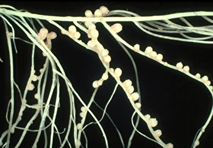
Rhizobium leguminosarum root nodules on the pea plant Pisum sativum. Mag: X 1.25

Background imageMicrobe Collection: Pink nodules of Rhizobium leguminosarum

Pink nodules of Rhizobium leguminosarum
Macrophotograph of pink nodules of the nitrogen- fixing bacteria Rhizobium leguminosarum on the roots of a pea plant (Pisum sativum)

Background imageMicrobe Collection: Streptococcus pneumoniae bacteria, SEM

Streptococcus pneumoniae bacteria, SEM
Streptococcus pneumoniae, coloured scanning electron micrograph (SEM). S. pneumoniae are oval, Gram-positive bacteria. They are part of the normal human bacterial flora

Background imageMicrobe Collection: Streptococcus oral bacteria, TEM

Streptococcus oral bacteria, TEM
Streptococcus oral bacteria. Coloured transmission electron micrograph (TEM) of two Streptococcus sanguinis bacteria. They are undergoing nuclear division (mitosis)

Background imageMicrobe Collection: Streptococcus bacteria, SEM

Streptococcus bacteria, SEM
Streptococcus bacteria. Coloured scanning electron micrograph (SEM) of Streptococcus sp. bacteria. Streptococci are round Gram-positive bacteria

Background imageMicrobe Collection: Coloured TEM of Enterococcus sp. bacteria

Coloured TEM of Enterococcus sp. bacteria
Enterococcus sp. bacteria. Coloured transmission electron micrograph (TEM) of a section through Enterococcus sp. bacteria. These were formerly grouped with the Streptococcus sp. bacteria

Background imageMicrobe Collection: Virus, artwork

Virus, artwork
Virus, computer artwork. Viruses contain a core of genetic material, either DNA (deoxyribonucleic acid) or RNA (ribonucleic acid) surrounded by a protein coat, or capsid

Background imageMicrobe Collection: Bacterial culture

Bacterial culture. Petri dish containing bacterial cultures (red). The bacteria are grown on agar jelly (beige), which provides all the nutrients needed by bacteria for growth

Background imageMicrobe Collection: Virus particles, conceptual artwork

Virus particles, conceptual artwork
Virus particles, conceptual computer artwork. Viruses contain a core of genetic material, either DNA (deoxyribonucleic acid) or RNA (ribonucleic acid) surrounded by a protein coat, or capsid

Background imageMicrobe Collection: Stromatolites at Hamelin Pool, Shark Bay, western Australia. These are living representatives of

Stromatolites at Hamelin Pool, Shark Bay, western Australia. These are living representatives of the oldest living
ROG-13733 Stromatolites at Hamelin Pool, Shark Bay, western Australia These are living representatives of the oldest living organisms, over 3.5 billion years old as fossils

Background imageMicrobe Collection: Myxococcus xanthus bacteria, SEM

Myxococcus xanthus bacteria, SEM
Myxococcus xanthus bacteria, coloured scanningelectron micrograph (SEM). M. xanthus is a rodshaped Gram-negative bacterium that resides inthe soil

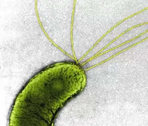
Background imageMicrobe Collection: Proteus vulgaris bacterium, TEM

Proteus vulgaris bacterium, TEM
Proteus vulgaris bacterium, colouredtransmission electron micrograph (TEM). The yellowthreads are flagella, hair-like structures thatthe bacterium beats for motility

Background imageMicrobe Collection: Oral bacteria, TEM

Oral bacteria, TEM
Oral bacteria. Computer-enhanced colouredtransmission electron micrograph (TEM) of Porphyromonas gingivalis bacteria (formerlynamed Bacteroides gingivalis )

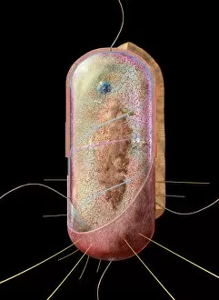
Background imageMicrobe Collection: Bacterial cell

Bacterial cell, computer artwork. This is ageneralised bacterium, illustrating features ofdifferent bacterial types that would not becombined in a single cell

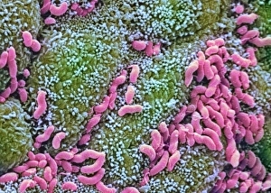
Background imageMicrobe Collection: Helicobacter pylori bacteria in stomach

Helicobacter pylori bacteria in stomach
Helicobacter pylori bacteria. Colouredscanning electron micrograph (SEM) of Helicobacter pylori bacteria (pink), a causeof gastritis (stomach lining inflammation)

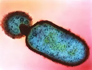
Background imageMicrobe Collection: Listeria monocytogenes bacteria, TEM

Listeria monocytogenes bacteria, TEM
Listeria bacteria. Coloured transmissionelectron micrograph (TEM) of a Listeria monocytogenes bacterium (centre to bottomright). It is dividing by a process of unequalcell growth known as budding

Background imageMicrobe Collection: Unidentified rod-shaped bacteria

Unidentified rod-shaped bacteria
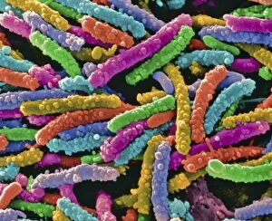
Bacteria. Coloured scanning electronmicrograph (SEM) of unidentified bacilli(rod-shaped bacteria). Magnification unknown

Background imageMicrobe Collection: Robert Koch in Kimberley

Robert Koch in Kimberley
HEINRICH HERMANN ROBERT KOCH German physician and pioneer bacteriologist in search of the Rinderpest microbe at Kimberley

Background imageMicrobe Collection: Futuristic study of microbes

Futuristic study of microbes
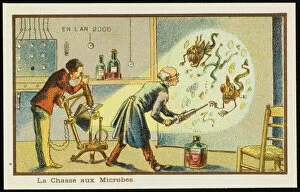
A scientist studies microbes magnified on a screen


All Professionally Made to Order for Quick Shipping

"Unveiling the Invisible: Exploring the Microbial World" In a realm unseen by the naked eye, lies a fascinating universe teeming with life. Meet the microbe, an extraordinary entity that encompasses various organisms and shapes our understanding of biology. HeLa cells, immortalized and widely used in research, have revolutionized medical science as they continue to unravel mysteries within our own bodies. Captured under a light microscope (C017 / 8299), their intricate structures reveal secrets waiting to be discovered. Witness the battle between neutrophils and MRSA as these microscopic warriors engage in a relentless struggle for survival. In an astonishing scanning electron microscope image (C018 / 8596), observe how these immune cells engulf harmful bacteria, showcasing nature's defense mechanisms at work. Eagerly multiplying like tiny soldiers on a mission, E. coli bacteria emerge into view through another SEM image. Their presence reminds us of both their beneficial role in digestion and potential harm when found in contaminated food or water sources. Salmonella bacteria take center stage next; their distinctive features magnified by yet another SEM image. These notorious culprits behind foodborne illnesses serve as reminders of proper hygiene practices necessary for safeguarding public health. Behold Yersinia pestis bacteria captured in vibrant colors through transmission electron microscopy (TEM). This captivating visual representation highlights the infamous pathogen responsible for devastating outbreaks such as the Black Death throughout history. Delving deeper into this microbial world reveals Candida fungus thriving amidst its surroundings. A striking SEM image showcases its filamentous structure – reminding us of its ability to cause infections if given favorable conditions. Returning to HeLa cells once more (light micrograph C017 / 8298), we are reminded of Henrietta Lacks' invaluable contribution to medical research - her immortal cell line continues to pave new paths towards scientific breakthroughs even decades after her passing.