Fraud Blocker Skip to main content

Microfilament Collection

Microfilaments, also known as actin filaments, play a crucial role in various cellular processes

Background imageMicrofilament Collection: HeLa cells, light micrograph C017 / 8299

HeLa cells, light micrograph C017 / 8299
HeLa cells, multiphoton fluorescence micrograph (MFM). The cell nuclei, which contain the cells genetic information, are purple. Microtubules are blue and actin microfilaments are red

Background imageMicrofilament Collection: Illustration of muscle contraction

Illustration of muscle contraction

Background imageMicrofilament Collection: Detail of deep back muscles with a close-up of sprain, strain and spasm

Detail of deep back muscles with a close-up of sprain, strain and spasm

Background imageMicrofilament Collection: Conceptual image of cytoskeleton

Conceptual image of cytoskeleton. The cytoskeleton is a cellular scaffolding or skeleton contained within a cells cytoplasm, and is present in all cells

Background imageMicrofilament Collection: Illustration of muscle contraction (with labels)

Illustration of muscle contraction (with labels)

Background imageMicrofilament Collection: Comparative illustration of plant and animal cell anatomy (with labels)

Comparative illustration of plant and animal cell anatomy (with labels)

Background imageMicrofilament Collection: Torn muscle fibers with healing stages surrounding

Torn muscle fibers with healing stages surrounding. 1. Macrophage clean-up. 2. New cells migrate to repair. 3. Cells differentiate to become muscle cells

Background imageMicrofilament Collection: Fibroblast cell, fluorescent micrograph

Fibroblast cell, fluorescent micrograph
Fibroblast cell. Confocal laser scanning micrograph of a fibroblast cell. The cell nucleus, which contains the cells genetic information, is green

Background imageMicrofilament Collection: Intracellular transport, artwork C013 / 5001

Intracellular transport, artwork C013 / 5001
Intracellular transport. Computer artwork of a vesicle (sphere) being transported along a microtubule (blue and green) by a kinesin motor protein (orange)

Background imageMicrofilament Collection: Intracellular transport, artwork C013 / 4997

Intracellular transport, artwork C013 / 4997
Intracellular transport. Computer artwork of vesicles (spheres) being transported from a Golgi body (blue, left) around the cell by microtubules (string-like)

Background imageMicrofilament Collection: Intracellular transport, artwork C013 / 4995

Intracellular transport, artwork C013 / 4995
Intracellular transport. Computer artwork of vesicles (spheres) being transported from a Golgi body (blue, top) around the cell by microtubules (string-like)


All Professionally Made to Order for Quick Shipping

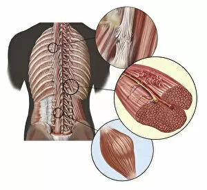
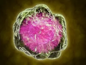
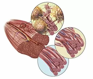
Microfilaments, also known as actin filaments, play a crucial role in various cellular processes. Derived from the protein actin, these thin and flexible structures are found in all eukaryotic cells. In HeLa cells, they can be visualized using a light micrograph C017 / 8299. The intricate network of these filaments provides structural support to the cell and helps maintain its shape. An illustration of muscle contraction showcases how microfilaments interact with myosin to generate movement. As myosin pulls on the actin filaments, muscles contract and enable us to perform physical activities. A close-up of sprain, strain, and spasm in deep back muscles reveals the importance integrity for proper muscle function. Any disruption or damage to these filaments can result in painful conditions that affect our mobility. Comparative illustrations of plant and animal cell anatomy highlight the presence of microfilaments in both types of cells. These filaments contribute to cell division, cytoplasmic streaming, and organelle movement. Conceptual images depicting the cytoskeleton emphasize how microfilaments form an essential part of this dynamic framework within cells. They provide mechanical strength while allowing flexibility for cellular movements. Labels on illustrations demonstrating muscle contraction and plant-animal cell anatomy help identify specific components involved with microfilament interactions. Torn muscle fibers surrounding healing stages remind us that repairing damaged tissues heavily relies on functional microfilament networks. Fibroblast cells play a key role in this process by producing new collagen fibers along the injury site. Fluorescent micrographs showcasing fibroblast cells further highlight their involvement in tissue repair. These specialized cells migrate towards injured areas where they secrete extracellular matrix components necessary for wound healing. Whether it's maintaining cell structure or facilitating muscle contractions and tissue repair processes like wound healing, microfilaments prove indispensable in various biological contexts.