
Microplicae Collection
Microplicae: Exploring the Intricate World of Fish Skin and Oesophagus Epithelium Delving into the microscopic realm, we uncover the fascinating world of microplicae


All Professionally Made to Order for Quick Shipping
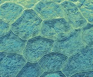
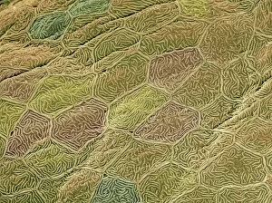
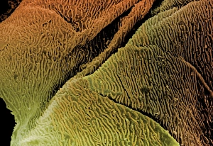
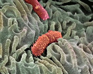
Microplicae: Exploring the Intricate World of Fish Skin and Oesophagus Epithelium Delving into the microscopic realm, we uncover the fascinating world of microplicae. These tiny structures, found in zebra fish skin and oesophagus epithelium, have captivated scientists with their intricate beauty. Using scanning electron microscopy (SEM), researchers have been able to capture stunning images that reveal the delicate nature of these microplicae. In zebra fish skin, SEM showcases a mesmerizing pattern of overlapping ridges, resembling an artistic masterpiece. Moving on to fish skin cell SEM images, we witness a closer look at individual cells forming this remarkable structure. The intricacy is astounding as each cell contributes to the overall strength and protection provided by microplicae. Shifting our focus towards oesophagus epithelium, SEM unveils another captivating sight. The lining of this vital organ displays a textured surface adorned with countless tiny projections known as microplicae. These structures play a crucial role in increasing surface area for efficient nutrient absorption and protection against harmful substances. Coloured SEM images further enhance our understanding of oesophagus epithelial cells' complexity. Vibrant hues highlight different types of cells working harmoniously together to maintain optimal functioning. As we delve deeper into oesophagus lining SEM visuals, it becomes evident how these minute details contribute to its functionality. Microplicae create an elaborate landscape that aids in digestion while safeguarding against potential damage or irritation. Exploring microplicae through SEM offers us glimpses into nature's intricate design within zebra fish skin and oesophageal tissues. These extraordinary structures remind us once again how even at the smallest scale, beauty can be found everywhere if we take the time to look closely enough.