Fraud Blocker Skip to main content

Microscope Collection (page 3)

"Unveiling the Unseen: Exploring the Microscopic World" Step into a realm of hidden wonders

Background imageMicroscope Collection: Microscope composed by Galileo Galilei and made by Giuseppe Campani, 1650-1700 (cardboard, wood)

Microscope composed by Galileo Galilei and made by Giuseppe Campani, 1650-1700 (cardboard, wood, metal and leather)
DEX5040791 Microscope composed by Galileo Galilei and made by Giuseppe Campani, 1650-1700 (cardboard, wood, metal and leather); Istituto e Museo di Storia della Scienza, Florence

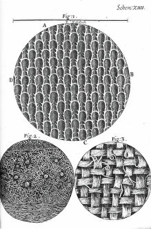
Background imageMicroscope Collection: Micrographia

Micrographia
2626672 Micrographia; (add.info.: Illustration from 'Micrographia' 1665; by Robert Hooke, 1635-1703. Hooke's observations through various lenses)

Background imageMicroscope Collection: Micrographia

Micrographia
2626679 Micrographia; (add.info.: Illustration from 'Micrographia' 1665; by Robert Hooke, 1635-1703. Hooke's observations through various lenses)

Background imageMicroscope Collection: France, Rhone-Alpes, Rhone (69), Lyon: laboratory of Monsieur Girard, 1904

France, Rhone-Alpes, Rhone (69), Lyon: laboratory of Monsieur Girard, 1904
LUX4792351 France, Rhone-Alpes, Rhone (69), Lyon: laboratory of Monsieur Girard, 1904; (add.info.: France, Rhone-Alpes, Rhone (69), Lyon: laboratory of Monsieur Girard

Background imageMicroscope Collection: Micrographia

Micrographia
2626675 Micrographia; (add.info.: Illustration from 'Micrographia' 1665; by Robert Hooke, 1635-1703. Hooke's observations through various lenses)

Background imageMicroscope Collection: Mikhail Alekseevich Pavlov During the Time of Wintering, 1912. Creator: Nikolay Vasilyevich Pinegin

Mikhail Alekseevich Pavlov During the Time of Wintering, 1912. Creator: Nikolay Vasilyevich Pinegin
Mikhail Alekseevich Pavlov During the Time of Wintering, 1912. From a collection consisting of an album and individual photographs from the family archive of geologist Mikhail Alekseevich Pavlov

Background imageMicroscope Collection: Four performances concerning the Nature and Medical Congress in Leiden, 1889

Four performances concerning the Nature and Medical Congress in Leiden, 1889. Creator: Theo van Hoytema
Four performances concerning the Nature and Medical Congress in Leiden, 1889. At the top a portrait of Prof. dr. Dr. G. van Overbeek de Meijer; At the bottom a portrait of Prof. dr. Dr. A.A.W
Background imageMicroscope Collection: Class in physiology, 1904. Creator: Frances Benjamin Johnston

Class in physiology, 1904. Creator: Frances Benjamin Johnston
Class in physiology, 1904

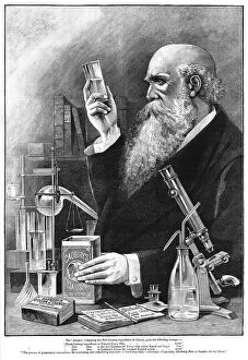
Background imageMicroscope Collection: 'Cadbury's Cocoa, 1890. Creator: Unknown

'Cadbury's Cocoa, 1890. Creator: Unknown
'Cadbury's Cocoa, 1890. From "The Graphic. An Illustrated Weekly Newspaper", Volume 42. July to December, 1890

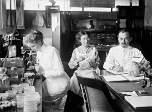
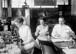
Background imageMicroscope Collection: Bacteriology - Dr. George Stiles, 1912. Creator: Harris & Ewing. Bacteriology - Dr

Bacteriology - Dr. George Stiles, 1912. Creator: Harris & Ewing. Bacteriology - Dr. George Stiles, 1912
Bacteriology - Dr. George Stiles, 1912. [Two women and man in laboratory - USA?]

Background imageMicroscope Collection: Study N°7 | Epidermis

Study N°7 | Epidermis
Thoth Adan

Background imageMicroscope Collection: Petri Dish

Petri Dish
Thoth Adan

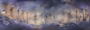
Background imageMicroscope Collection: Forest dreams

Forest dreams
Kristina Zvinakeviciute

Background imageMicroscope Collection: Focus stacked, inverted image of Diatoms on a microscope slide

Focus stacked, inverted image of Diatoms on a microscope slide. Diatoms are single-celled algae which produce approximately 25% of the oxygen we breathe as well as being responsible for approximately

Background imageMicroscope Collection: Female students of a medical school. Territory of Krasnoyarsk

Female students of a medical school. Territory of Krasnoyarsk Russia. Date of Photograph:1980 ca

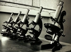
Background imageMicroscope Collection: Microscopes Koriska, School of Science in the Real Commercial Technical Institute F

Microscopes Koriska, School of Science in the Real Commercial Technical Institute F. Besta in Treviso Treviso. Date of Photograph:1950-1960

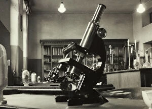
Background imageMicroscope Collection: Microscope Koriska, School of Science or Merceology Museum in the Real Commercial Technical

Microscope Koriska, School of Science or Merceology Museum in the Real Commercial Technical Institute F. Besta in Treviso Treviso. Date of Photograph:1950-1960

Background imageMicroscope Collection: Laboratory of Merceology at the Technical Institute of Bolzano; on the counter some microscopes

Laboratory of Merceology at the Technical Institute of Bolzano; on the counter some microscopes and a measuring instrument for flour moisture measurement Bolzano. Date of Photograph:1930-1940

Background imageMicroscope Collection: Women in charge of inspecting vials

Women in charge of inspecting vials Italy. Date of Photograph:1955 ca

Background imageMicroscope Collection: Vertical microscope dating back to the eighteenth century. The picture was taken during

Vertical microscope dating back to the eighteenth century. The picture was taken during the Exhibition of Science History in 1929, in Florence. Florence Exhibition of Science History

Background imageMicroscope Collection: The image shows cattotropic microscopes of the scientist Giovan Battists Amici

The image shows cattotropic microscopes of the scientist Giovan Battists Amici, with drawers and accessories, in the foreground is an ocular micrometer, with double images

Background imageMicroscope Collection: English vertical microscope dating back to the twentieth century belonging to the Carbonelli

English vertical microscope dating back to the twentieth century belonging to the Carbonelli Collection, Rome. The picture was taken during the Exhibition of Science History in 1929

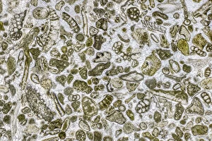
Background imageMicroscope Collection: Limestone microfossils in a 0. 03mm-thick slice of fossil-rich limestone viewed at high

Limestone microfossils in a 0. 03mm-thick slice of fossil-rich limestone viewed at high magnification
Limestone microfossils in a 0.03mm-thick slice of fossil-rich limestone viewed at high magnification, image area is 3mm across in real life

Background imageMicroscope Collection: J Lancaster & Son, Colmore Row, Birmingham

J Lancaster & Son, Colmore Row, Birmingham
J Lancaster & Son, Cameras, Photographic and Scientific Equipment, Colmore Row, Birmingham. circa 1900

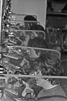
Background imageMicroscope Collection: Women night workers at Texas Instruments Ltd in Bedford Circa February 1970

Women night workers at Texas Instruments Ltd in Bedford Circa February 1970. Workers seen here grading transistors and integrated circuit boards

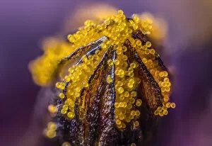
Background imageMicroscope Collection: African daisy (Osteospermum jucundum) at approx 10x magnification

African daisy (Osteospermum jucundum) at approx 10x magnification, composite flower showing individual florets full of golden pollen grains, Monmouthshire, Wales, UK, July

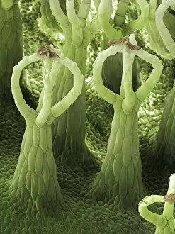
Background imageMicroscope Collection: Salvinia Effect of Salvinia natans

Salvinia Effect of Salvinia natans
Scanning electron micrograph of leaf detail of Salvinia natans, a floating fern type plant which has superhydrophobic trichomes or hairs on the upper surface of its leaves

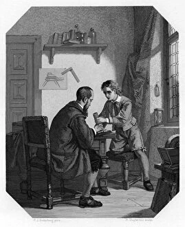
Background imageMicroscope Collection: Hans Jansen and his son Sacharias, c1870. Artist: H Sluyter

Hans Jansen and his son Sacharias, c1870. Artist: H Sluyter
Hans Jansen and his son Sacharias, c1870. Some historians credit Sacharias Jansen, a Middelburg spectacle maker, with the invention of the telescope and the microscope

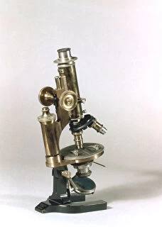
Background imageMicroscope Collection: Microscope made by C Reichert, Vienna, 1895. Artist: C Reichert

Microscope made by C Reichert, Vienna, 1895. Artist: C Reichert
Microscope made by C Reichert, Vienna, 1895. The microscope featured objectives of different powers which could be turned into viewing position as required

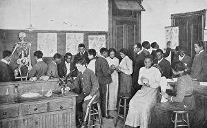
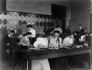
Background imageMicroscope Collection: Students in a science class using microscopes, Western High School, Washington, D.C. (1899?)

Students in a science class using microscopes, Western High School, Washington, D.C. (1899?)

Background imageMicroscope Collection: Physiology Laboratory, London School of Medicine for Women

Physiology Laboratory, London School of Medicine for Women, Hunter Street. Date: 1898

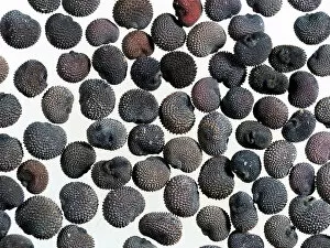
Background imageMicroscope Collection: Red campion (Silene dioica) seeds, microscopic view

Red campion (Silene dioica) seeds, microscopic view

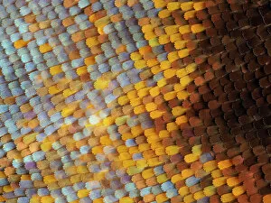
Background imageMicroscope Collection: Abstract close-up of butterfly wing scales

Abstract close-up of butterfly wing scales

Background imageMicroscope Collection: Microscope by the Zeiss company from Jena

Microscope by the Zeiss company from Jena
7277941 Microscope by the Zeiss company from Jena by Unknown Artist, (19th century); Private Collection; (add.info.: Microscope by the Zeiss company from Jena)

Background imageMicroscope Collection: False-coloured scanning electron micrograph of a Thrip's (Thysanoptera) head

False-coloured scanning electron micrograph of a Thrip's (Thysanoptera) head, the bulging compound eyes can be seen on either side of the head

Background imageMicroscope Collection: Soil bacteria cultured on nutrient agar from a soil sample collected in Via Gellia Woods, Derbyshire

Soil bacteria cultured on nutrient agar from a soil sample collected in Via Gellia Woods, Derbyshire. Image size in real life 10mm across. UK. March

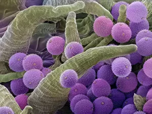
Background imageMicroscope Collection: Pollen grains on the style of a Harebell (Campanula rotundifolia), Hoe Grange Nature Reserve

Pollen grains on the style of a Harebell (Campanula rotundifolia), Hoe Grange Nature Reserve, Derbyshire, UK. August

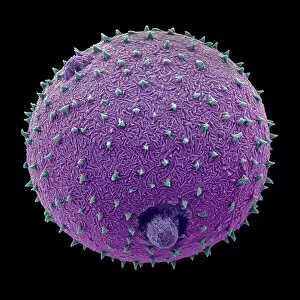
Background imageMicroscope Collection: Harebell (Campanula rotundifolia) pollen grain, false-coloured Scanning Electron Micrograph

Harebell (Campanula rotundifolia) pollen grain, false-coloured Scanning Electron Micrograph

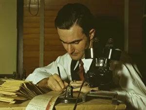
Background imageMicroscope Collection: Geologist examining cuttings from wildcat well, Amarillo, Texas, (1943?). Creator: John Vachon

Geologist examining cuttings from wildcat well, Amarillo, Texas, (1943?). Creator: John Vachon
Geologist examining cuttings from wildcat well, Amarillo, Texas

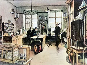
Background imageMicroscope Collection: Portrait of Robert Koch in his laboratory (engraving, 19th century)

Portrait of Robert Koch in his laboratory (engraving, 19th century)
LRI4670026 Portrait of Robert Koch in his laboratory (engraving, 19th century) by Unknown Artist, (19th century); Private Collection; (add.info)

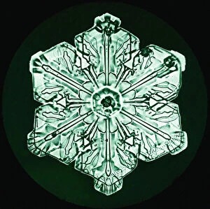
Background imageMicroscope Collection: Study showing the form and structure of a Snowflake, c. 1902 (photo)

Study showing the form and structure of a Snowflake, c. 1902 (photo)
7252346 Study showing the form and structure of a Snowflake, c.1902 (photo) by Bentley, Wilson A. (1865-1931); Private Collection; (add.info.: Wilson Alwyn Bentley (1865 - 1931)

Background imageMicroscope Collection: Study showing the form and structure of a Snowflake, c. 1902 (photo)

Study showing the form and structure of a Snowflake, c. 1902 (photo)
7252352 Study showing the form and structure of a Snowflake, c.1902 (photo) by Bentley, Wilson A. (1865-1931); Private Collection; (add.info.: Wilson Alwyn Bentley (1865 - 1931)

Background imageMicroscope Collection: Study showing the form and structure of a Snowflake, c. 1902 (photo)

Study showing the form and structure of a Snowflake, c. 1902 (photo)
7252343 Study showing the form and structure of a Snowflake, c.1902 (photo) by Bentley, Wilson A. (1865-1931); Private Collection; (add.info.: Wilson Alwyn Bentley (1865 - 1931)

Background imageMicroscope Collection: Study showing the form and structure of a Snowflake, c. 1902 (photo)

Study showing the form and structure of a Snowflake, c. 1902 (photo)
7252353 Study showing the form and structure of a Snowflake, c.1902 (photo) by Bentley, Wilson A. (1865-1931); Private Collection; (add.info.: Wilson Alwyn Bentley (1865 - 1931)

Background imageMicroscope Collection: Study showing the form and structure of a Snowflake, c. 1902 (photo)

Study showing the form and structure of a Snowflake, c. 1902 (photo)
7252357 Study showing the form and structure of a Snowflake, c.1902 (photo) by Bentley, Wilson A. (1865-1931); Private Collection; (add.info.: Wilson Alwyn Bentley (1865 - 1931)

Background imageMicroscope Collection: Study of Snowflakes, c. 1903 (photo)

Study of Snowflakes, c. 1903 (photo)
7252363 Study of Snowflakes, c.1903 (photo) by Bentley, Wilson A. (1865-1931); Private Collection; (add.info.: Wilson Alwyn Bentley (1865 - 1931), also known as Snowflake Bentley

Background imageMicroscope Collection: Study showing the form and structure of a Snowflake, c. 1902 (photo)

Study showing the form and structure of a Snowflake, c. 1902 (photo)
7252351 Study showing the form and structure of a Snowflake, c.1902 (photo) by Bentley, Wilson A. (1865-1931); Private Collection; (add.info.: Wilson Alwyn Bentley (1865 - 1931)


All Professionally Made to Order for Quick Shipping

"Unveiling the Unseen: Exploring the Microscopic World" Step into a realm of hidden wonders, where scientists like Rosalind Franklin and Santiago Ramon y Cajal have unraveled the mysteries through their microscopic lens. From Liver Fluke to Norovirus particles, these pioneers have paved the way for groundbreaking discoveries. Witness the intricate beauty of Snail teeth and Fat cells as they come alive under the microscope's gaze. Marvel at Lubbock's Graphic 84, a visual masterpiece capturing the essence of this microcosmic universe. Delve deeper into this microscopic world and encounter Plasmodium sp. , a malarial parasite that has plagued humanity for centuries. Explore E. Coli bacterium in all its glory, revealing both its potential dangers and scientific significance. Through microscope slide preparation techniques, we gain insight into complex organisms like never before. Discover how Fleming used his photographic skills to document crucial moments in microbiology history. The microscope acts as our window into an unseen dimension - it allows us to explore realms beyond our naked eye's reach, and is through this powerful tool that we continue to unravel nature's secrets one magnified image at a time. So join us on this journey "Under the Microscope, " where every slide holds a story waiting to be told – stories that shape our understanding of life itself.