Fraud Blocker Skip to main content

Mitosis Collection

Mitosis, the incredible process of cell division, can be observed under a light micrograph

Background imageMitosis Collection: Mitosis, light micrograph

Mitosis, light micrograph
Mitosis. Confocal light micrograph of the stages of mitosis (nuclear division) and cytokinesis (cell division). During mitosis the nuclear envelope disintegrates (3rd image) and the chromosomes (blue)

Background imageMitosis Collection: Cell division, fluorescent micrograph

Cell division, fluorescent micrograph
Cell division. Immunofluorescent light micrograph of a human epithelial cell (centre) during the late anaphase stage of mitosis

Background imageMitosis Collection: Embryo development 24-36 hours after fertilization

Embryo development 24-36 hours after fertilization

Background imageMitosis Collection: Dividing cells

Dividing cells. Computer artwork of a spiral of cells during mitosis (nuclear division). During mitosis two daughter nuclei are formed from one parent nucleus

Background imageMitosis Collection: Cell division, fluorescent micrograph

Cell division, fluorescent micrograph
Cell division. Immunofluorescent light micrograph of a human epithelial cell (centre) during the interphase stage of mitosis

Background imageMitosis Collection: Blastocyst, artwork F006 / 2190

Blastocyst, artwork F006 / 2190
Blastocyst. Computer artwork of a 100-cell blastocyst embryo

Background imageMitosis Collection: Growth

Growth
Thoth Adan

Background imageMitosis Collection: Nuclear division, illustration

Nuclear division, illustration
Nuclear division, computer illustration

Background imageMitosis Collection: Plant cell mitosis, light micrograph

Plant cell mitosis, light micrograph
Plant cell mitosis. Light micrograph of root tip cells from an onion (Allium sp.) undergoing cell division (mitosis). From top left to bottom right

Background imageMitosis Collection: Illustration of Atlantic Salmon (Salmo salar), male fish swimming in ocean

Illustration of Atlantic Salmon (Salmo salar), male fish swimming in ocean

Background imageMitosis Collection: Zygote, artwork F006 / 2563

Zygote, artwork F006 / 2563
Zygote. Computer artwork of a fertilised ovum (egg), or zygote

Background imageMitosis Collection: Diagram of human egg being fertilized in the fallopian tube

Diagram of human egg being fertilized in the fallopian tube

Background imageMitosis Collection: Cross section biomedical illustration of mitosis where nucleus membrane form around each set of

Cross section biomedical illustration of mitosis where nucleus membrane form around each set of chromosomes and the cell begins to divide in two

Background imageMitosis Collection: Cross section biomedical illustration of mitosis with new cells form

Cross section biomedical illustration of mitosis with new cells form, each having a central nucleus containing identical set of chromosomes

Background imageMitosis Collection: Cluster of blastomeres forming a developing morula

Cluster of blastomeres forming a developing morula (early stage of embryonic development)

Background imageMitosis Collection: Microscopic view of a stem cell

Microscopic view of a stem cell. Stem cells are biological cells found in all multicellular organisms that can divide through mitosis and differentiate into diverse specialized cell types

Background imageMitosis Collection: Zygote development 24-36 hours after fertilization

Zygote development 24-36 hours after fertilization

Background imageMitosis Collection: Microscopic view of stem cell development

Microscopic view of stem cell development. In adult organisms, stem cells and progenitor cells act as a repair system for the body, replenishing adult tissues

Background imageMitosis Collection: Conceptual image of mitosis

Conceptual image of mitosis. Mitosis is the process in the cell cycle by which a cell duplicates into two genetically identical sets of chromosomes

Background imageMitosis Collection: Microscopic view of cell division

Microscopic view of cell division

Background imageMitosis Collection: Picture No. 10852165

Picture No. 10852165
Broad Bean Root Tip - shows cell division / mitosis (Vicia faba) Date:

Background imageMitosis Collection: Blastocyst embryo, light micrograph F008 / 3573

Blastocyst embryo, light micrograph F008 / 3573
Blastocyst embryo, light micrograph. This embryo is five to six days old

Background imageMitosis Collection: Zygote, artwork F006 / 2564

Zygote, artwork F006 / 2564
Zygote. Computer artwork of a fertilised ovum (egg), or zygote

Background imageMitosis Collection: Morula embryo, artwork F006 / 2398

Morula embryo, artwork F006 / 2398
Morula embryo. Computer artwork of a 16-cell morula embryo

Background imageMitosis Collection: Eight-cell embryo, artwork F006 / 2200

Eight-cell embryo, artwork F006 / 2200
Eight-cell embryo, computer artwork

Background imageMitosis Collection: Implanted blastocyst, artwork F006 / 2199

Implanted blastocyst, artwork F006 / 2199
Implanted blastocyst. Computer artwork of a blastocyst embryo implanted in the wall of the uterus (womb)

Background imageMitosis Collection: Blastocyst, artwork F006 / 2197

Blastocyst, artwork F006 / 2197
Blastocyst. Computer artwork of a 100-cell blastocyst embryo

Background imageMitosis Collection: Implanted blastocyst, artwork F006 / 2194

Implanted blastocyst, artwork F006 / 2194
Implanted blastocyst. Computer artwork of a blastocyst embryo implanted in the wall of the uterus (womb)

Background imageMitosis Collection: Blastocyst, artwork F006 / 2198

Blastocyst, artwork F006 / 2198
Blastocyst. Computer artwork of a 58-cell blastocyst embryo

Background imageMitosis Collection: Blastocyst, artwork F006 / 2192

Blastocyst, artwork F006 / 2192
Blastocyst. Computer artwork of a 100-cell blastocyst embryo

Background imageMitosis Collection: Implanted blastocyst, artwork F006 / 2189

Implanted blastocyst, artwork F006 / 2189
Implanted blastocyst. Computer artwork of a blastocyst embryo implanted in the wall of the uterus (womb)

Background imageMitosis Collection: Eight-cell embryo, artwork F006 / 2170

Eight-cell embryo, artwork F006 / 2170
Morula embryo. Computer artwork of a 16-cell morula embryo

Background imageMitosis Collection: Four-cell embryo, artwork F006 / 2169

Four-cell embryo, artwork F006 / 2169
Four-cell embryo, computer artwork

Background imageMitosis Collection: Two-cell embryo, artwork F006 / 2167

Two-cell embryo, artwork F006 / 2167
Two-cell embryo, computer artwork

Background imageMitosis Collection: Cell division, artwork F006 / 9891

Cell division, artwork F006 / 9891
Cell division, computer artwork

Background imageMitosis Collection: Cell division, artwork F006 / 9892

Cell division, artwork F006 / 9892
Cell division, computer artwork

Background imageMitosis Collection: Roundworm germ cells, light micrograph C016 / 9538

Roundworm germ cells, light micrograph C016 / 9538
Roundworm germ cells. Light micrograph of germ cells from a roundworm (Ascaris sp.), undergoing mitosis (nuclear division)

Background imageMitosis Collection: Chromosome segregation protein

Chromosome segregation protein, molecular model. This proteins function is to aid the process of chromosome segregation during cell division and replication

Background imageMitosis Collection: Dividing cell, TEM

Dividing cell, TEM
Dividing cell. Transmission electron micrograph (TEM) of a section through a cell dividing through the process of mitosis

Background imageMitosis Collection: Cellular polarity, illustration C018 / 0775

Cellular polarity, illustration C018 / 0775
Cellular Polarity. Conceptual illustration of cell polarity research and its medical implications

Background imageMitosis Collection: Dividing cancer cell, SEM C014 / 0362

Dividing cancer cell, SEM C014 / 0362
Dividing cancer cell. Coloured scanning electron micrograph (SEM) of a colorectal cancer cell undergoing mitosis (nuclear division) and splitting into two daughter cells (left and right)

Background imageMitosis Collection: Mouth cancer cell dividing, SEM

Mouth cancer cell dividing, SEM
Mouth cancer dividing. Coloured scanning electron micrograph (SEM) of a squamous cell carcinoma (cancer) cell from a human mouth undergoing mitosis (nuclear division)

Background imageMitosis Collection: Dividing liver cancer cell, SEM

Dividing liver cancer cell, SEM
Dividing liver cancer cell. Coloured scanning electron micrograph (SEM) of a hepatocellular carcinoma (HCC) cell undergoing mitosis (nuclear division)

Background imageMitosis Collection: Dividing cancer cell, SEM C014 / 0361

Dividing cancer cell, SEM C014 / 0361
Dividing cancer cell. Coloured scanning electron micrograph (SEM) of a colorectal cancer cell undergoing mitosis (nuclear division) and splitting into two daughter cells (left and right)

Background imageMitosis Collection: Dividing bone cancer cell, SEM

Dividing bone cancer cell, SEM
SEM Dividing Osteosarcoma cells Osteosarcoma is an aggressive malignant neoplasm arising from primitive transformed cells of mesenchymal origin (and thus a sarcoma)

Background imageMitosis Collection: Lung cancer cell division, artwork C013 / 4630

Lung cancer cell division, artwork C013 / 4630
Lung cancer cell division. Computer artwork of a lung cancer cell during cell division (cytokinesis). The two daughter cells remain temporarily joined by a cytoplasmic bridge (centre)

Background imageMitosis Collection: Mitosis in root tip of Broad Bean, LM

Mitosis in root tip of Broad Bean, LM
Light micrograph (longitudinal section) of the cells in the root tip of a broad bean plant Vicia faba. The dark spot in the centre of each cell is the nucleus

Background imageMitosis Collection: 4-cell embryo embryo, artwork

4-cell embryo embryo, artwork
4-cell embryo. Image 3 of 4. Computer model representing 4 daughter cells. The development of an embryo is called embryogenesis


All Professionally Made to Order for Quick Shipping

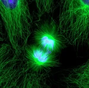
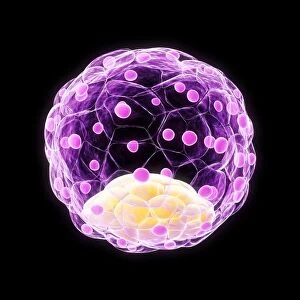
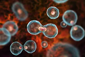
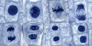
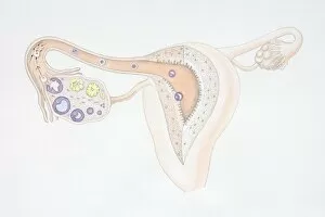
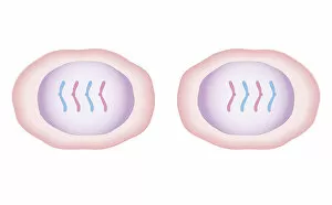
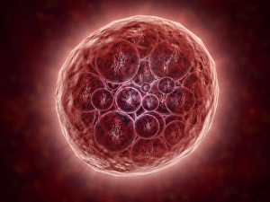
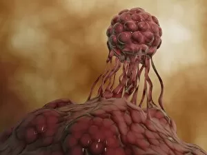
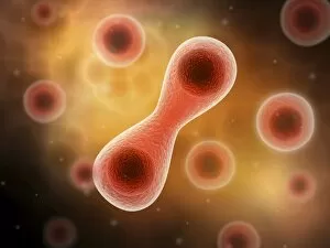
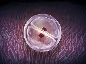
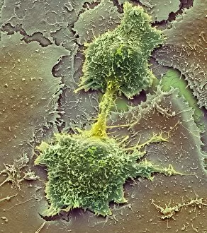
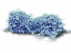
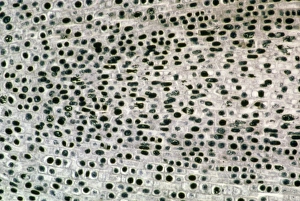
Mitosis, the incredible process of cell division, can be observed under a light micrograph. The fluorescent micrograph showcases the intricate details of this fundamental biological event. In embryo development, just 24-36 hours after fertilization, dividing cells undergo mitosis to ensure proper growth and formation. The blastocyst, depicted in artwork F006/2190, represents the early stages of development where mitotic divisions are crucial for shaping an organism's future. Through nuclear division illustrated vividly, we witness how chromosomes align on threads that form across the cell as the nucleus breaks down. Not limited to animal cells alone, plant cell it is captured in a stunning light micrograph. This emphasizes that mitosis plays a vital role in all living organisms' growth and reproduction. Even Atlantic Salmon (Salmo salar), male fish swimming gracefully in the ocean waters benefit from this remarkable process. Mitosis ensures their bodies regenerate and grow efficiently throughout their lives. In human reproduction, a diagram illustrates how a human egg is fertilized within the fallopian tube. It highlights how mitotic divisions occur during early embryonic development to create new life. Cross-section biomedical illustrations provide us with an intimate view at its core – showcasing how nuclei break down while threads form across cells with chromosomes lining up meticulously along these threads. From microscopic observations to artistic representations and scientific diagrams - each depiction reminds us of the significance in every aspect of life's existence: from single-celled organisms to complex beings like humans.