
Molecular Layer Collection
The molecular layer, a vital component of the cerebellum structure, is a fascinating sight under the microscope


All Professionally Made to Order for Quick Shipping
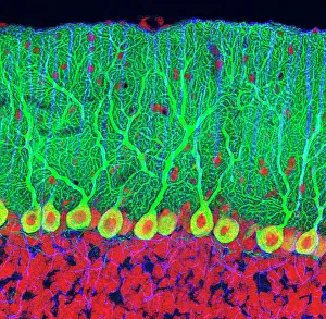
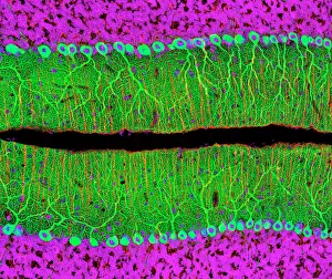
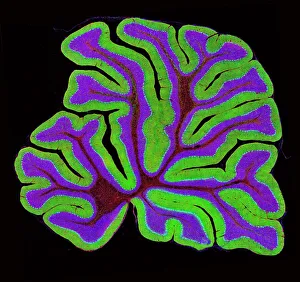
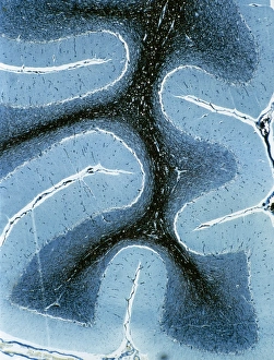
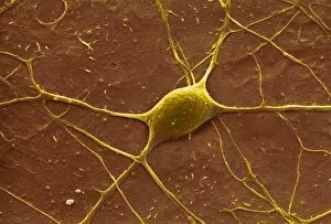
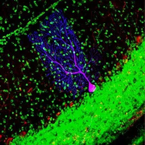
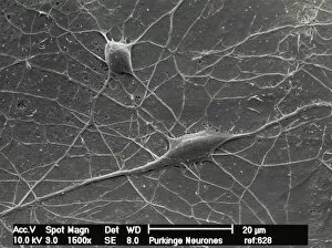
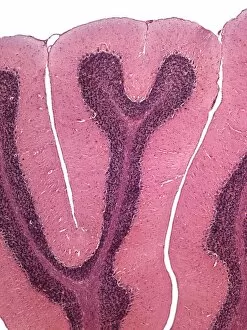
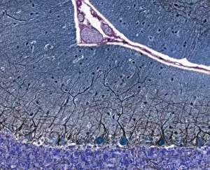
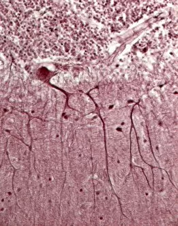
The molecular layer, a vital component of the cerebellum structure, is a fascinating sight under the microscope. This intricate network of Purkinje nerve cells in the cerebellum forms an awe-inspiring tapestry that plays a crucial role in our motor coordination and balance. When observed through a light micrograph, the complexity and beauty of this layer become apparent. The delicate arrangement of Purkinje nerve cells can be seen intertwining with each other, creating an intricate web-like pattern. These specialized neurons are responsible for transmitting information from various parts of the brain to coordinate smooth movements and maintain equilibrium. As we delve deeper into the cerebellum tissue using light microscopy, we witness more stunning details within its structure. The layers appear distinct yet interconnected, forming an organized architecture that supports its essential functions. In one particular image captured by a light micrograph, we observe both cortex and medulla regions of the cerebellum side by side. The contrast between these two areas highlights their unique characteristics while emphasizing their interdependence in maintaining proper motor control. Another striking feature revealed through these microscopic images is the presence of numerous Purkinje nerve cells scattered throughout the tissue. Their elongated bodies extend like branches across this molecular layer, showcasing their significance in relaying signals necessary for precise movement execution. A closer look at these individual Purkinje nerve cells unveils even more intricacies when viewed under high magnification using scanning electron microscopy (SEM). The detailed surface features come alive as if inviting us to explore further into their world. In summary, exploring the molecular layer provides us with captivating glimpses into how our brains orchestrate coordinated movements effortlessly. Through careful examination using different imaging techniques such as light micrographs and SEMs, scientists continue to unravel its complexities and unlock new insights into neurological disorders affecting motor skills.