Fraud Blocker Skip to main content

Molecular Model Collection

Molecular models offer a glimpse into the intricate world of science and medicine, revealing the hidden secrets of life at a microscopic level

Background imageMolecular Model Collection: DNA transcription, molecular model

DNA transcription, molecular model. Secondary structure of the enzyme RNA polymerase II synthesising a mRNA (messenger ribonucleic acid, lilac) strand from a DNA (deoxyribonucleic acid)

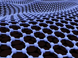
Background imageMolecular Model Collection: Graphene sheet, artwork C016 / 8274

Graphene sheet, artwork C016 / 8274
Graphene sheet. Computer artwork showing the molecular structure of a graphene sheet. Graphene is a single layer of graphite

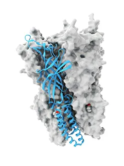
Background imageMolecular Model Collection: Anaesthetic inhibiting an ion channel C015 / 6718

Anaesthetic inhibiting an ion channel C015 / 6718
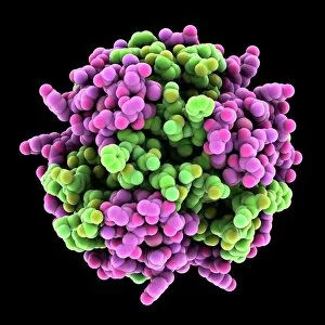
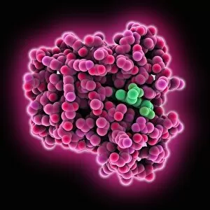
Anaesthetic inhibiting an ion channel. Computer model showing the structure of propofol anaesthetic drug molecules (spheres)

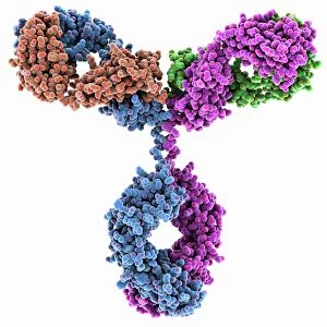
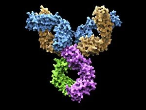
Background imageMolecular Model Collection: Immunoglobulin G antibody molecule

Immunoglobulin G antibody molecule. Computer model of the secondary structure of immunoglobulin G (IgG). This is the most abundant immunoglobulin and is found in all body fluids
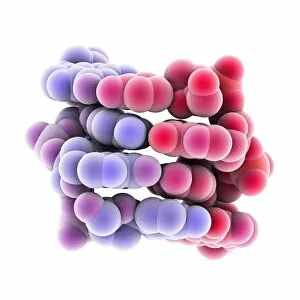
Background imageMolecular Model Collection: Double-stranded RNA molecule

Double-stranded RNA molecule. Computer model of the structure of double-stranded RNA (ribonucleic acid). The majority of RNA in a cell is in the single-stranded form

Background imageMolecular Model Collection: Immunoglobulin G antibody molecule F007 / 9894

Immunoglobulin G antibody molecule F007 / 9894
Immunoglobulin G antibody molecule. Computer model of the secondary structure of immunoglobulin G (IgG). This is the most abundant immunoglobulin and is found in all body fluids

Background imageMolecular Model Collection: 2C-B psychedelic drug, molecular model

2C-B psychedelic drug, molecular model. Atoms are represented as spheres and are colour-coded: carbon (grey), hydrogen (white), oxygen (pink), nitrogen (blue) and bromine (dark red)

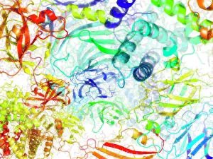
Background imageMolecular Model Collection: Secondary structure of proteins, artwork

Secondary structure of proteins, artwork
Secondary structure of proteins, computer artwork. The secondary structure is the shape taken by the strands of proteins, which are biological polymers of amino acids

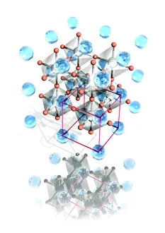
Background imageMolecular Model Collection: Perovskite crystal structure

Perovskite crystal structure. Perovskite is the name for the mineral calcium titanium oxide (CaTiO3). It forms a crystal structure that is very common in oxide minerals

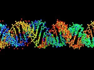
Background imageMolecular Model Collection: DNA molecule, computer model

DNA molecule, computer model
DNA molecule. Computer artwork of the molecular structure of DNA (deoxyribonucleic acid). The DNA molecule is composed of two strands twisted into a double helix

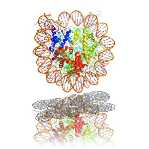
Background imageMolecular Model Collection: Nucleosome molecule

Nucleosome molecule, computer model. A nucleosome is a subunit of chromatin, the substance that forms chromosomes. It consists of a short length of DNA (deoxyribonucleic acid)

Background imageMolecular Model Collection: DNA nucleosome, molecular model

DNA nucleosome, molecular model
DNA nucleosome. Molecular model of a nucleosome, the fundamental repeating unit used to package DNA (deoxyribonucleic acid) inside cell nuclei

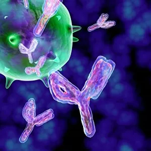
Background imageMolecular Model Collection: Antibodies, artwork

Antibodies, artwork
Computer artwork of antibody molecules showing the structure of an immunoglobulin G (IgG) molecule. This is the most abundant immunoglobulin and is found in all body fluids

Background imageMolecular Model Collection: Caffeine drug molecule

Caffeine drug molecule
Caffeine. Computer model of a molecule of the alkaloid, stimulant and legal drug caffeine. Caffeine is most often consumed in drinks like tea and coffee

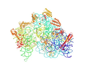
Background imageMolecular Model Collection: Bacterial ribosome

Bacterial ribosome. Computer model showing the secondary structure of a 30S (small) ribosomal sub-unit from the bacteria Thermus thermophilus

Background imageMolecular Model Collection: HIV reverse transcription enzyme

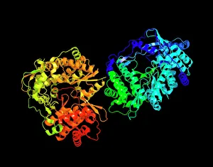
HIV reverse transcription enzyme. Molecular models of the reverse transcriptase enzyme found in HIV (the human immunodeficiency virus)

Background imageMolecular Model Collection: Hepatitis C virus enzyme, molecular model

Hepatitis C virus enzyme, molecular model
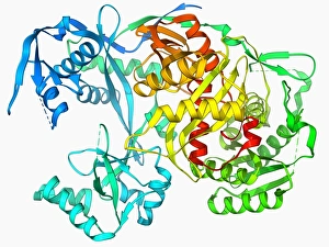
Hepatitis C virus enzyme. Molecular model of a genetic enzyme from the Hepatitis C virus. This enzyme is called HC-J4 RNA polymerase

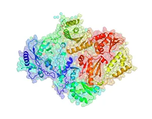
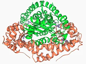
Background imageMolecular Model Collection: Glutamine synthetase enzyme

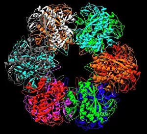
Glutamine synthetase enzyme computer model. This is a ligase enzyme, which forms chemical bonds between molecules. The different colours show the different subunits that comprise the protein

Background imageMolecular Model Collection: Insulin molecule, artwork

Insulin molecule, artwork
Artwork of a molecule of human insulin, a hormone produced by the pancreas which controls levels of glucose in the blood. The molecule consists of two peptide chains

Background imageMolecular Model Collection: Vitamin B12, molecular model

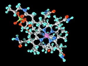
Vitamin B12, molecular model. Vitamin B12 (cyanocobalamin) is an essential nutrient that humans are unable to produce and need to obtain from their diet

Background imageMolecular Model Collection: RNA-editing enzyme, molecular model

RNA-editing enzyme, molecular model
RNA-editing enzyme. Molecular model of a left-handed, RNA double helix (Z-RNA, centre) bound by the Z alpha domain of the human RNA-editing enzyme ADAR1 (double-stranded RNA adenosine deaminase)

Background imageMolecular Model Collection: Zinc fingers bound to a DNA strand

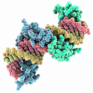
Zinc fingers bound to a DNA strand, molecular model. The double helix of DNA (deoxyribonucleic acid, red and yellow) is seen here with two Zif268 proteins (blue and green)

Background imageMolecular Model Collection: Myoglobin molecule C015 / 5702

Myoglobin molecule C015 / 5702
Myoglobin molecule. Computer model showing the structure of a myoglobin molecule. Myoglobin is a protein found in muscle tissue

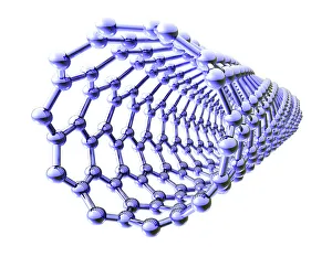
Background imageMolecular Model Collection: Carbon nanotube

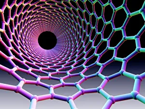
Carbon nanotube. Computer artwork showing the hexagonal carbon structure of a nanotube, or buckytube

Background imageMolecular Model Collection: Capsaicin molecule

Capsaicin molecule
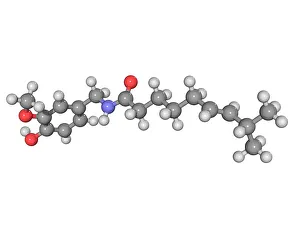
Capsaicin, molecular model. This chemical gives chilies their heat and causes a burning sensation when ingested. It is a secondary metabolite of the chili plant (Capsicum sp)

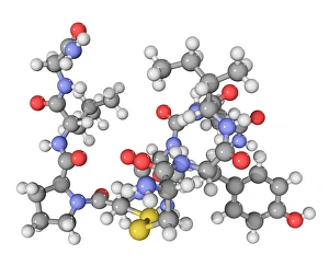
Background imageMolecular Model Collection: Oxytocin neurotransmitter molecule

Oxytocin neurotransmitter molecule. Computer model showing the structure of the neurotransmitter and hormone Oxytocin. Atoms are colour-coded spheres (carbon: dark grey, hydrogen: light grey)

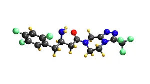
Background imageMolecular Model Collection: Januvia diabetes drug molecule

Januvia diabetes drug molecule
Januvia diabetes drug, molecular model. Januvia (sitagliptin) is a hypoglycaemic drug, one that reduces blood sugar levels

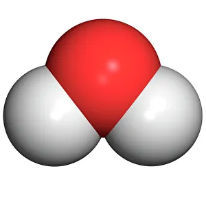
Background imageMolecular Model Collection: Water molecule

Water molecule. Computer model of a molecule of water. Atoms are colour-coded: oxygen (red) and hydrogen (white). Water is one of the most abundant chemicals on Earth

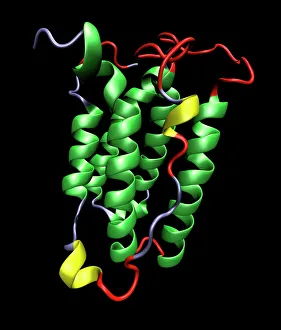
Background imageMolecular Model Collection: Interferon molecule

Interferon molecule. Computer model showing the secondary structure of a molecule of interferon. Interferons are proteins produced by white blood cells as part of the immune response to invading

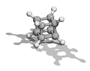
Background imageMolecular Model Collection: Cubane molecule

Cubane molecule. Computer model showing the structure of a molecule of cubane (C8H8). Atoms are represented as colour-coded spheres (carbon, grey; hydrogen)

Background imageMolecular Model Collection: Manganese superoxide dismutase enzyme F006 / 9423

Manganese superoxide dismutase enzyme F006 / 9423
Manganese superoxide dismutase enzyme, molecular model. This enzyme scavenges and decomposes the potentially toxic first reduction product, superoxide, of aerobic respiration

Background imageMolecular Model Collection: SARS coronavirus protein

SARS coronavirus protein. Molecular model of the ORF-9b protein produced by the SARS (severe acute respiratory syndrome) coronavirus

Background imageMolecular Model Collection: Cytochrome b5 molecule C015 / 6696

Cytochrome b5 molecule C015 / 6696
Cytochrome b5. Molecular model of cytochrome b5 from a cows liver. Cytochrome molecules perform oxidation and reduction reactions for electron transport

Background imageMolecular Model Collection: Z-DNA tetramer molecule C015 / 6557

Z-DNA tetramer molecule C015 / 6557
Z-DNA (deoxyribonucleic acid) tetramer, molecular model. DNA is composed of two strands twisted into a double helix. This is a tetramer of the molecule, containing four strands

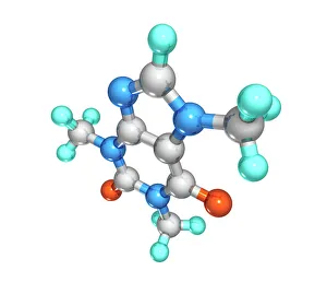
Background imageMolecular Model Collection: Caffeine, molecular model

Caffeine, molecular model. Atoms are represented as spheres and are colour-coded: carbon (grey), hydrogen (green), oxygen (red) and nitrogen (blue)

Background imageMolecular Model Collection: Aflatoxin, molecular model

Aflatoxin, molecular model. Atoms are represented as spheres and are colour-coded: carbon (grey), hydrogen (white) and oxygen (red)

Background imageMolecular Model Collection: Rotavirus particle, artwork

Rotavirus particle, artwork
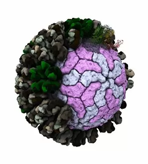
Rotavirus particle. Cut-away artwork showing the structure of the rotavirus icosahedral capsid (protein coat). The capsid, which encloses the RNA (ribonucleic acid) genome

Background imageMolecular Model Collection: Cholera toxin, molecular model

Cholera toxin, molecular model
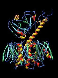
Cholera toxin. Molecular model of the secondary structure of cholera enterotoxin (intestinal toxin). The molecule consists of two subunits, A (top) and B (bottom)

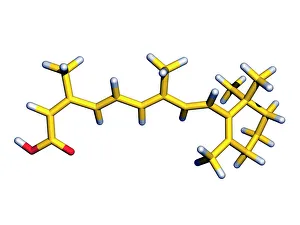
Background imageMolecular Model Collection: Isotretinoin anti-acne drug

Isotretinoin anti-acne drug, molecular model. Atoms are represented as tubes and are colour- coded; carbon (yellow), hydrogen (white) and oxygen (red)

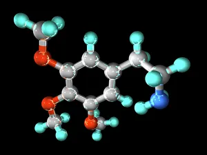
Background imageMolecular Model Collection: Mescaline hallucinogenic drug molecule

Mescaline hallucinogenic drug molecule
Mescaline hallucinogenic drug, molecular model. Mescaline is a hallucinogenic drug, produced from the dried tops (buttons) of the peyote cactus (Lophophora williamsii)

Background imageMolecular Model Collection: Nanotube technology, computer artwork

Nanotube technology, computer artwork
Nanotube technology. Computer artwork of a cylindrical fullerene molecule (carbon nanotube). The hexagonal carbon structure of the nanotube is shown here

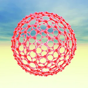
Background imageMolecular Model Collection: Fullerene molecule, computer artwork

Fullerene molecule, computer artwork
Fullerene molecule. Computer artwork of the spherical fullerene molecule C320. Fullerenes are a structural type (allotrope) of carbon

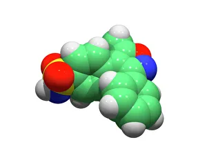
Background imageMolecular Model Collection: Valdecoxib anti-inflammatory drug

Valdecoxib anti-inflammatory drug
Valdecoxib, computer model. This drug was used in the treatment of osteoarthritis, rheumatoid arthritis and menstrual symptoms under the trade name Bextra

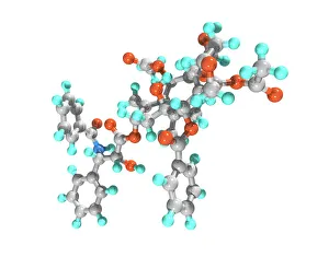
Background imageMolecular Model Collection: Paclitaxel drug molecule

Paclitaxel drug molecule
Paclitaxel. Computer model of a molecule of the drug paclitaxel. It is sold under the brand name Taxol. It is a chemotherapy drug, used to treat cancers

Background imageMolecular Model Collection: DNA strands, illustration

DNA strands, illustration
DNA strands. Computer illustration showing the structure of double stranded DNA (deoxyribonucleic acid) molecules. DNA is composed of two strands twisted into a double helix

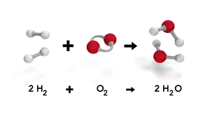
Background imageMolecular Model Collection: Reaction of hydrogen and oxygen to water C017 / 3598

Reaction of hydrogen and oxygen to water C017 / 3598
Reaction of hydrogen and oxygen to water. Computer artwork of a balanced chemical equation showing how two hydrogen (H2, white) molecules (left) combine with a single oxygen (O2)

Background imageMolecular Model Collection: Argonaute protein molecule F006 / 9526

Argonaute protein molecule F006 / 9526
Argonaute protein, molecular model. This protein forms the RNA-induced silencing complex (RISC) along with a small interfering RNA (ribonucleic acid) molecule

Background imageMolecular Model Collection: TFAM transcription factor bound to DNA C015 / 7059

TFAM transcription factor bound to DNA C015 / 7059
TFAM transcription factor bound to DNA, molecular model. Human mitochondrial transcription factor A (TFAM, green) bound to a strand of DNA (deoxyribonucleic acid, blue and pink)


All Professionally Made to Order for Quick Shipping

Molecular models offer a glimpse into the intricate world of science and medicine, revealing the hidden secrets of life at a microscopic level. In one captivating image, an anaesthetic molecule is seen inhibiting an ion channel C015/6718, unlocking new possibilities for pain management. Another striking model showcases the complex structure of a double-stranded RNA molecule, shedding light on its crucial role in gene regulation and viral defense mechanisms. Delving deeper into genetics, we explore DNA transcription through a mesmerizing molecular model that unravels the intricate process of genetic information transfer. The spotlight then shifts to Immunoglobulin G antibody molecules - powerful defenders against pathogens - as their elegant structures are unveiled with precision. From F007/9894 variant to artwork-inspired representations, these models showcase the diversity within our immune system's arsenal. Venturing beyond traditional boundaries, we encounter 2C-B psychedelic drug's molecular model – offering insights into its unique chemical composition and potential therapeutic applications. Art meets science once again as we marvel at an artistic interpretation showcasing secondary structures of proteins; highlighting their vital roles in cellular functions. Inorganic wonders take center stage with the perovskite crystal structure model – unveiling its remarkable properties that revolutionize solar energy technology. Returning to genetics, we witness a computer-generated DNA molecule model providing us with invaluable insights into our blueprint for life. The complexity continues with the intricately designed nucleosome molecule – unraveling how DNA is packaged within our cells' nucleus while maintaining accessibility for essential processes. Finally, awe-inspiring artwork captures antibodies' beauty and significance as they stand tall against invading antigens. These captivating molecular models serve as windows into worlds unseen by the naked eye; bridging gaps between scientific exploration and artistic expression. They inspire curiosity and ignite imagination while propelling breakthroughs in fields ranging from medicine to materials science.