Fraud Blocker Skip to main content

Molecular Structure Collection (page 9)

"Molecular Structure: Unlocking the Secrets of Life's Building Blocks" From anaesthetics inhibiting ion channels to antidepressant molecules

Background imageMolecular Structure Collection: Acetylcholine molecule

Acetylcholine molecule
Acetylcholine, molecular model. Organic compound, neurotransmitter in nervous systems of many organisms. Atoms are represented as spheres and are colour-coded: carbon (grey), hydrogen (green-blue)

Background imageMolecular Structure Collection: Glutamic acid molecule

Glutamic acid molecule
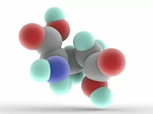
Glutamic acid, molecular model. Non-essential amino-acid. Important neurotransmitter. Atoms are represented as spheres and are colour-coded: carbon (grey), hydrogen (blue-green)

Background imageMolecular Structure Collection: Glutamine molecule

Glutamine molecule
Proline, molecular model. Non-essential alpha-amino acid, one of the 20 DNA-encoded amino acids. Atoms are represented as spheres and are colour-coded: carbon (grey), hydrogen (blue-green)

Background imageMolecular Structure Collection: Isoleucine molecule

Isoleucine molecule
Isoleucine, molecular model. Essential alpha-amino acid contained in eggs, soy protein, seaweed, turkey, chicken, lamb, cheese, and fish

Background imageMolecular Structure Collection: Threonine molecule

Threonine molecule
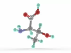
Threonine, molecular model. Essential alpha-amino acid and one of the 20 proteinogenic amino acids. Atoms are represented as spheres and are colour-coded: carbon (grey), hydrogen (blue-green)

Background imageMolecular Structure Collection: Gamma-aminobutyric acid GABA molecule

Gamma-aminobutyric acid GABA molecule
Gamma-aminobutyric acid (GABA), molecular model. Main inhibitory neurotransmitter in the central nervous system of mammalians

Background imageMolecular Structure Collection: Valine molecule

Valine molecule
Valine, molecular model. Essential alpha-amino acid and one of the 20 proteinogenic amino acids. Atoms are represented as spheres and are colour-coded: carbon (grey), hydrogen (blue-green)

Background imageMolecular Structure Collection: Phenylalanine molecule

Phenylalanine molecule
Phenylalanine, molecular model. Essential alpha-amino acid, one of the 20 common amino acids used to form proteins. Atoms are represented as spheres and are colour-coded: carbon (grey)

Background imageMolecular Structure Collection: Tryptophan molecule

Tryptophan molecule
Tryptophan, molecular model. Essential amino acid and one of the 20 standard amino acids. Atoms are represented as spheres and are colour-coded: carbon (grey), hydrogen (blue-green)

Background imageMolecular Structure Collection: Methionine molecule

Methionine molecule
Methionine, molecular model. Essential alpha-amino acid. Atoms are represented as spheres and are colour-coded: carbon (grey), hydrogen (blue-green), nitrogen (blue), oxygen (red) and sulfur (yellow)

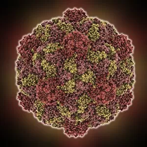
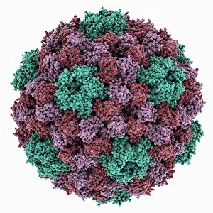
Background imageMolecular Structure Collection: HK97 bacteriophage capsid

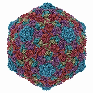
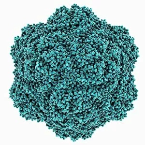
HK97 bacteriophage capsid, molecular model. Bacteriophages are viruses that infect bacteria, in this case enterobacteria such as E. coli (Escherichia coli), with the phage head shown here

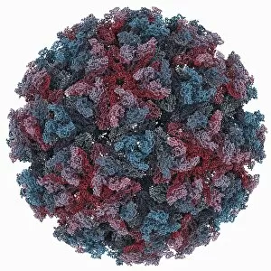
Background imageMolecular Structure Collection: Chikungunya virus capsid

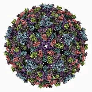
Chikungunya virus capsid, molecular model. This virus, transmitted by mosquitoes in tropical Africa and Asia, causes fever and joint pain in humans, similar to dengue fever

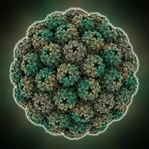
Background imageMolecular Structure Collection: HK97 bacteriophage procapsid

HK97 bacteriophage procapsid. Molecular model showing the structure of the prohead-I procapsid of the HK97 bacteriophage. Bacteriophages are viruses that infect bacteria

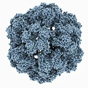
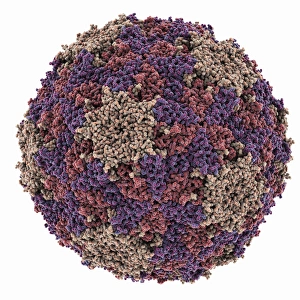
Background imageMolecular Structure Collection: Turnip yellow mosaic virus capsid

Turnip yellow mosaic virus capsid, molecular model. This virus infects a wide variety of plants, including crops such as turnips and cabbages, causing yellow patches on the leaves
Background imageMolecular Structure Collection: Sindbis virus capsid, molecular model

Sindbis virus capsid, molecular model. This virus, transmitted by mosquitoes, causes sindbis fever in humans. In viruses, the capsid is the protein shell that encloses the genetic material

Background imageMolecular Structure Collection: Murine polyomavirus capsid

Murine polyomavirus capsid, molecular model. This virus, one of a range named for their potential to cause multiple tumours, infects mice

Background imageMolecular Structure Collection: Brome mosaic virus capsid

Brome mosaic virus capsid, molecular model. This plant virus infects grasses, especially brome grasses, and also barley. It causes mosaic patches of discolouration

Background imageMolecular Structure Collection: Cowpea chlorotic mottle virus capsid

Cowpea chlorotic mottle virus capsid, molecular model. This virus (CCMV) infects the cowpea plant (Vigna unguiculata), causing yellow spots of discolouration

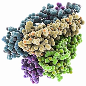
Background imageMolecular Structure Collection: Potassium ion channel protein structure

Potassium ion channel protein structure. Molecular model of a KcsA potassium ion (K+) channel from Streptomyces lividans bacteria

Background imageMolecular Structure Collection: Potassium ion channel beta subunit

Potassium ion channel beta subunit. Molecular model showing the structure a beta subunit of a voltage-dependent potassium (K+) channel

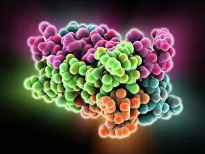
Background imageMolecular Structure Collection: KCNQ ion channel protein structure

KCNQ ion channel protein structure. Molecular model showing the protein structure of an ion channel domain. Ion channels are membrane-spanning proteins that form a pathway for the movement of

Background imageMolecular Structure Collection: Potassium ion channel cavity structure

Potassium ion channel cavity structure. Molecular model showing the structure of a cavity formed by potassium ion channel proteins

Background imageMolecular Structure Collection: Avian polyomavirus capsid

Avian polyomavirus capsid, molecular model. This virus, one of a range named for their potential to cause multiple tumours, infects birds. Discovered in budgerigars in 1981, it is often fatal

Background imageMolecular Structure Collection: Cytoplasmic polyhedrosis virus capsid

Cytoplasmic polyhedrosis virus capsid, molecular model. Part of the Cypovirus genus and invariably fatal, this insect virus is transmitted by contamination of leaves eaten (examples include silkworms)

Background imageMolecular Structure Collection: DNA molecule, artwork F007 / 1996

DNA molecule, artwork F007 / 1996
DNA molecule, computer artwork

Background imageMolecular Structure Collection: DNA molecule, artwork F007 / 1994

DNA molecule, artwork F007 / 1994
DNA molecule, computer artwork

Background imageMolecular Structure Collection: DNA molecule, artwork F007 / 1995

DNA molecule, artwork F007 / 1995
DNA molecule, computer artwork

Background imageMolecular Structure Collection: DNA molecule, artwork F007 / 1991

DNA molecule, artwork F007 / 1991
DNA molecule, computer artwork

Background imageMolecular Structure Collection: DNA molecule, artwork F007 / 1992

DNA molecule, artwork F007 / 1992
DNA molecule, computer artwork

Background imageMolecular Structure Collection: Theilers encephalomyelitis virus capsid

Theilers encephalomyelitis virus capsid, molecular model. This virus, which causes brain and spinal cord inflammation in mice, is used in research

Background imageMolecular Structure Collection: Lomitapide hypercholesterolemia drug F007 / 0160

Lomitapide hypercholesterolemia drug F007 / 0160
Lomitapide hypercholesterolemia drug, molecular model. Lomitapide is used in the treatment of homozygous familial hypercholesterolemia

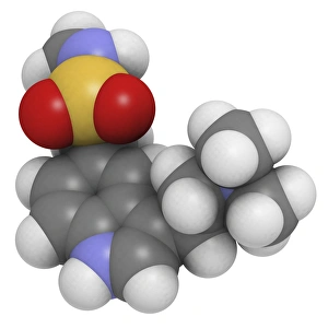
Background imageMolecular Structure Collection: Vortioxetine antidepressant drug F007 / 0207

Vortioxetine antidepressant drug F007 / 0207
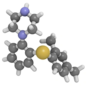
Vortioxetine antidepressant drug, molecular model. Atoms are represented as spheres and are colour-coded: hydrogen (white), carbon (grey), oxygen (red), sulfur (yellow) and nitrogen (blue)

Background imageMolecular Structure Collection: Vortioxetine antidepressant drug F007 / 0206

Vortioxetine antidepressant drug F007 / 0206
Vortioxetine antidepressant drug, molecular model. Atoms are represented as spheres and are colour-coded: hydrogen (white), carbon (grey), oxygen (red), sulfur (yellow) and nitrogen (blue)

Background imageMolecular Structure Collection: Tobacco necrosis virus capsid

Tobacco necrosis virus capsid, molecular model. This plant virus infects a wide rage of plants, including the tobacco plant for which it is named. The virus causes tissue death (necrosis)

Background imageMolecular Structure Collection: Vortioxetine antidepressant drug F007 / 0205

Vortioxetine antidepressant drug F007 / 0205
Vortioxetine antidepressant drug, molecular model. Atoms are represented as spheres and are colour-coded: hydrogen (white), carbon (grey), oxygen (red), sulfur (yellow) and nitrogen (blue)

Background imageMolecular Structure Collection: Vemurafenib melanoma drug F007 / 0204

Vemurafenib melanoma drug F007 / 0204
Vemurafenib melanoma drug, molecular model. Vemurafenib is a B-Raf enzyme inhibitor used in the treatment of melanoma skin cancer

Background imageMolecular Structure Collection: Vemurafenib melanoma drug F007 / 0203

Vemurafenib melanoma drug F007 / 0203
Vemurafenib melanoma drug, molecular model. Vemurafenib is a B-Raf enzyme inhibitor used in the treatment of melanoma skin cancer

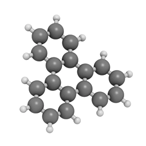
Background imageMolecular Structure Collection: Triphenylene hydrocarbon molecule F007 / 0202

Triphenylene hydrocarbon molecule F007 / 0202
Triphenylene polycyclic aromatic hydrocarbon (PAH), molecular model. Triphenylene is an environmental pollutant and suspected to be carcinogenic, mutagenic and teratogenic

Background imageMolecular Structure Collection: Triphenylene hydrocarbon molecule F007 / 0201

Triphenylene hydrocarbon molecule F007 / 0201
Triphenylene polycyclic aromatic hydrocarbon (PAH), molecular model. Triphenylene is an environmental pollutant and suspected to be carcinogenic, mutagenic and teratogenic

Background imageMolecular Structure Collection: Treprostinil drug, molecular model F007 / 0200

Treprostinil drug, molecular model F007 / 0200
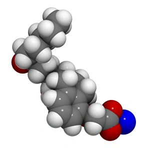
Treprostinil drug, molecular model. Treprostinil is used in the treatment of pulmonary arterial hypertension. Atoms are represented as spheres and are colour-coded: hydrogen (white), carbon (grey)

Background imageMolecular Structure Collection: Treprostinil drug, molecular model F007 / 0199

Treprostinil drug, molecular model F007 / 0199
Treprostinil drug, molecular model. Treprostinil is used in the treatment of pulmonary arterial hypertension. Atoms are represented as spheres and are colour-coded: hydrogen (white), carbon (grey)

Background imageMolecular Structure Collection: Trametinib melanoma cancer drug F007 / 0198

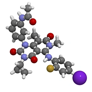
Trametinib melanoma cancer drug F007 / 0198
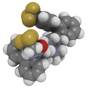
Trametinib melanoma cancer drug, molecular model. Atoms are represented as spheres and are colour-coded: hydrogen (white), carbon (grey), oxygen (red), fluorine (dark yellow)

Background imageMolecular Structure Collection: Trametinib melanoma cancer drug F007 / 0197

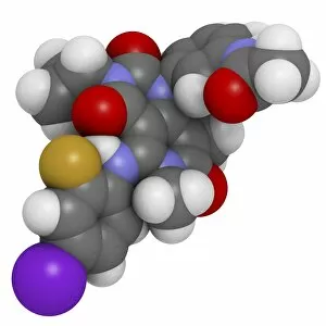
Trametinib melanoma cancer drug F007 / 0197
Trametinib melanoma cancer drug, molecular model. Atoms are represented as spheres and are colour-coded: hydrogen (white), carbon (grey), oxygen (red), fluorine (dark yellow)

Background imageMolecular Structure Collection: Tofacitinib rheumatoid arthritis drug F007 / 0196

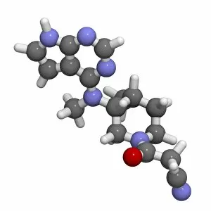
Tofacitinib rheumatoid arthritis drug F007 / 0196
Tofacitinib rheumatoid arthritis drug, molecular model. Atoms are represented as spheres and are colour-coded: hydrogen (white), carbon (grey), oxygen (red) and nitrogen (blue)

Background imageMolecular Structure Collection: Tofacitinib rheumatoid arthritis drug F007 / 0195

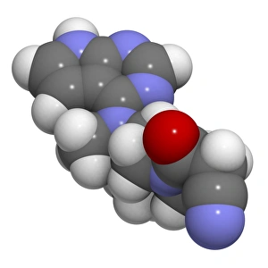
Tofacitinib rheumatoid arthritis drug F007 / 0195
Tofacitinib rheumatoid arthritis drug, molecular model. Atoms are represented as spheres and are colour-coded: hydrogen (white), carbon (grey), oxygen (red) and nitrogen (blue)

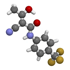
Background imageMolecular Structure Collection: Teriflunomide multiple sclerosis drug F007 / 0194

Teriflunomide multiple sclerosis drug F007 / 0194
Teriflunomide multiple sclerosis drug, molecular model. Atoms are represented as spheres and are colour-coded: hydrogen (white), carbon (grey), oxygen (red), fluorine (dark yellow) and nitrogen (blue)

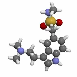
Background imageMolecular Structure Collection: Sumatriptan migraine headache drug F007 / 0192

Sumatriptan migraine headache drug F007 / 0192
Sumatriptan migraine headache drug, molecular model. Atoms are represented as spheres and are colour-coded: hydrogen (white), carbon (grey), oxygen (red), sulfur (yellow) and nitrogen (blue)

Background imageMolecular Structure Collection: Sumatriptan migraine headache drug F007 / 0190

Sumatriptan migraine headache drug F007 / 0190
Sumatriptan migraine headache drug, molecular model. Atoms are represented as spheres and are colour-coded: hydrogen (white), carbon (grey), oxygen (red), sulfur (yellow) and nitrogen (blue)


All Professionally Made to Order for Quick Shipping

"Molecular Structure: Unlocking the Secrets of Life's Building Blocks" From anaesthetics inhibiting ion channels to antidepressant molecules, the intricate world holds endless wonders. The C015 / 6718 anaesthetic molecule delicately interacts with ion channels, altering their function and providing relief from pain. Meanwhile, Amitriptyline, an antidepressant molecule, works its magic by modulating neurotransmitters in our brains. In the realm of immunity, Immunoglobulin G antibody F007 / 9894 stands tall as a defender against pathogens. Its unique structure allows it to recognize and neutralize foreign invaders effectively. On another front, DNA artwork showcases the elegance and complexity that underlies all life forms on Earth. Creatine amino acid molecule fuels our muscles during intense physical activities while nanotube technology revolutionizes various industries with its exceptional properties. These tiny tubes hold immense potential for advancements in medicine and materials science alike. Zinc fingers bound to a DNA strand demonstrate how proteins can precisely interact with genetic material. This interaction plays a crucial role in gene regulation and expression. Carbon nanotubes take center stage once again as they exhibit remarkable strength and conductivity at the nano-scale level. Oxytocin neurotransmitter molecule reminds us of love's powerful influence on human connections—its presence promotes bonding between individuals. Manganese superoxide dismutase enzyme F006 / 9423 safeguards our cells by combating harmful free radicals that contribute to aging and disease. Even viruses have their own molecular structures; SARS coronavirus protein represents one such example—a key player in viral replication within host cells. Conceptual artwork further explores nanotube technology's limitless possibilities—the fusion of imagination and scientific innovation knows no bounds here. As we delve deeper into understanding molecular structures, we unravel nature's blueprint for life itself—one atom at a time. These captivating glimpses into the microscopic world remind us of both the fragility and resilience found within the building blocks of existence.