
Mycobacterium Tuberculosis Collection
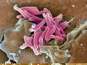
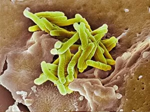
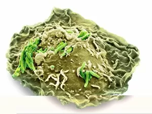
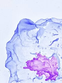
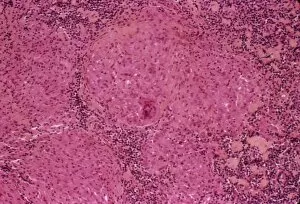
"Mycobacterium tuberculosis: A Stealthy Intruder Unveiled" Tuberculosis bacteria, also known as Mycobacterium tuberculosis


All Professionally Made to Order for Quick Shipping
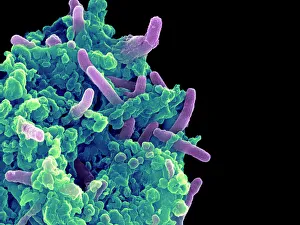
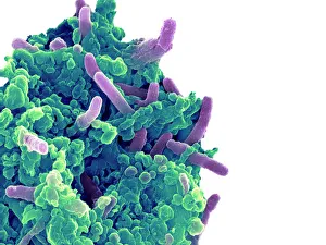
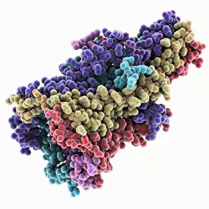
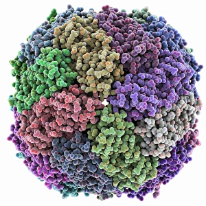
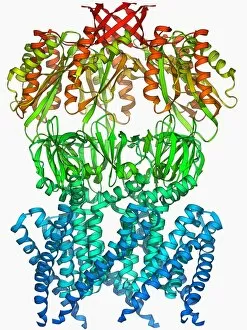
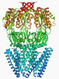
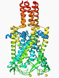
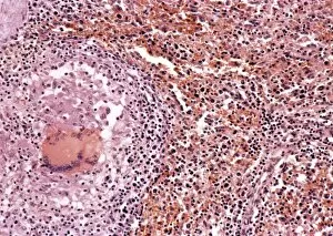
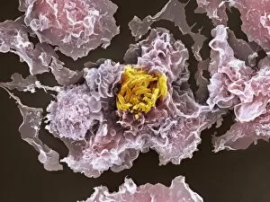
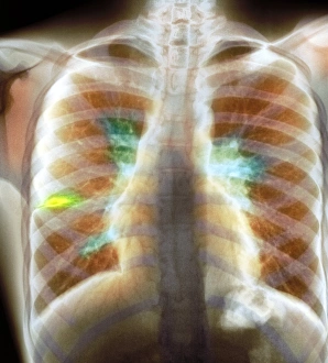
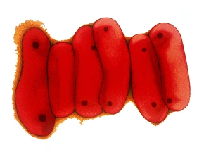
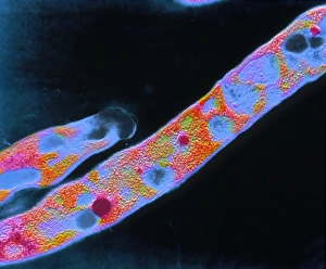
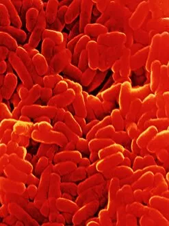
"Mycobacterium tuberculosis: A Stealthy Intruder Unveiled" Tuberculosis bacteria, also known as Mycobacterium tuberculosis, are responsible for causing the infectious disease tuberculosis. This captivating scanning electron microscope (SEM) image reveals the moment when these tiny bacteria infect a macrophage, a type of immune cell. Witness the intense battle between the macrophage and TB bacteria as it engulfs them in this mesmerizing SEM capture. Delving deeper into their interaction, this SEM image showcases how TB bacteria invade and infect a macrophage, evading our body's defense mechanisms. An artistic representation highlights its unique structure and features that make it such an elusive pathogen. Behold the relentless pursuit of a macrophage engulfing TB bacteria captured through high-resolution SEM imaging - a true testament to our body's defense system at work. Another striking SEM image unravels the intricate details of how TB bacteria infiltrate and colonize within a macrophage, leading to infection and disease progression. Explore the fascinating structure of MscL ion channel protein - an essential component in understanding how Mycobacterium tuberculosis manipulates host cells during infection. Remembering Robert Koch - the brilliant German bacteriologist who made groundbreaking discoveries about Mycobacterium tuberculosis and laid foundations for modern diagnostics and treatment strategies against tuberculosis. Dive into molecular modeling with this depiction of an iron-containing protein associated with Mycobacterium tuberculosis - shedding light on its role in bacterial survival within human hosts. Further unraveling mysteries at the molecular level, explore MscL ion channel protein structure - providing insights into potential targets for novel drug development against Mycobacterium tuberculosis infections. Discover another key player in cellular response to stress caused by TB infection: MscS ion channel protein structure F006/9650.