
Myelination Collection
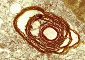
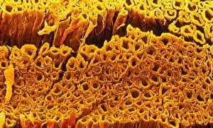
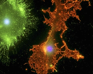
Myelination: Unveiling the Intricacies of Nerve Fibres In the microscopic realm, a fascinating process known as myelination takes place within our bodies


All Professionally Made to Order for Quick Shipping
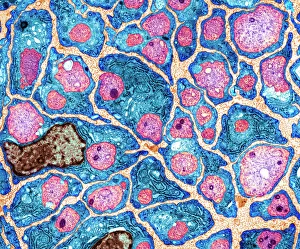
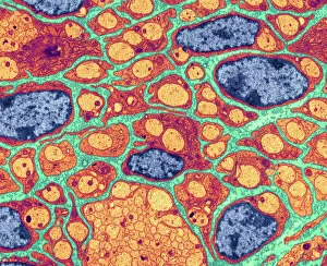
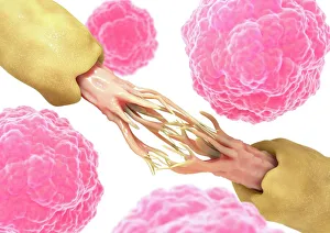
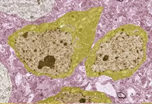
Myelination: Unveiling the Intricacies of Nerve Fibres In the microscopic realm, a fascinating process known as myelination takes place within our bodies. Through the lens of a transmission electron microscope (TEM), we can witness this intricate phenomenon unfold. The TEM captures stunning images showcasing myelination of nerve fibres in all its glory. These snapshots reveal the delicate and complex web formed by these specialized cells, known as oligodendrocytes. They meticulously wrap themselves around nerve axons, creating a protective sheath called myelin. As we delve deeper into this microscopic world, we stumble upon another captivating artwork depicting nerve damage and stem cells. It serves as a reminder that although myelin plays an essential role in maintaining proper neuronal function, it is not invincible to harm or degradation. Examining further TEM images, we encounter brain cells - the building blocks of our thoughts and actions. Each image showcases their unique structures and interconnectedness. The complexity becomes evident as countless brain cells intertwine with one another like an intricately woven tapestry. Amongst these brain cells lies an extraordinary network of nerves responsible for transmitting information throughout our body. The TEM reveals how myelin surrounds each nerve axon like a protective shield, ensuring efficient signal propagation from one cell to another. These glimpses into the world beneath our skin shed light on both the beauty and fragility of our nervous system's architecture. Myelination holds immense importance in preserving neural integrity while facilitating rapid communication between different regions of our body. Understanding this process allows us to appreciate how crucial it is for maintaining optimal cognitive function and motor skills. Moreover, it highlights potential avenues for research aimed at developing therapies that harness stem cells' regenerative power to repair damaged nerves. Through mesmerizing TEM imagery capturing myelination's wonders alongside breathtaking depictions of brain cells and nerve networks – we gain insight into the remarkable intricacies of our nervous system.