
Myological Collection
"Myological Marvels: Exploring the Intricacies of Muscles Through Art and Science" In this captivating collection


All Professionally Made to Order for Quick Shipping
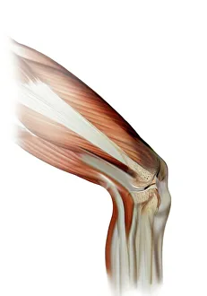
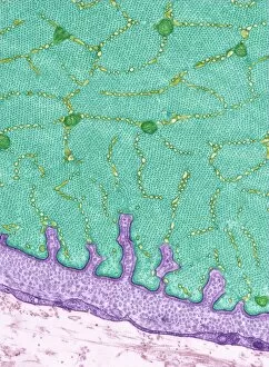
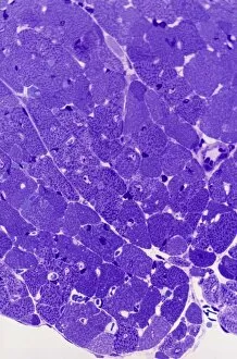
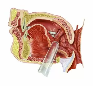
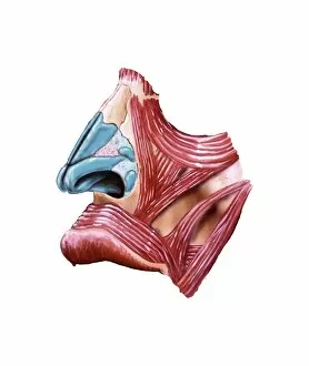
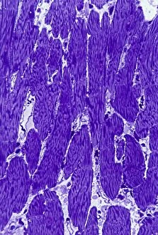
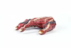
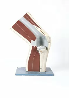
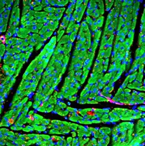
"Myological Marvels: Exploring the Intricacies of Muscles Through Art and Science" In this captivating collection, we delve into the fascinating world of myology - the study of muscles. From damaged knee ligaments to intricate eye muscles, each artwork tells a unique story about our body's remarkable strength and resilience. The first piece draws us in with its depiction of a damaged knee ligament. The artist skillfully captures the fragility and pain associated with such an injury, reminding us of the importance of taking care of our joints. Moving on, we encounter TEM C014 / 1468 - a mesmerizing portrayal of eye muscles. These intricate structures are responsible for our ability to see and convey emotions through subtle movements. This artwork serves as a reminder that even the smallest muscle can have a profound impact on our daily lives. Next, we come across stunning illustrations showcasing shoulder muscles and upper arm muscles. These artworks highlight their power and versatility, emphasizing their role in everyday activities like lifting objects or reaching out for a hug. As we continue our journey through myology, we stumble upon a breathtaking light micrograph capturing cardiac muscle cells in all their glory. The delicate striations reveal the incredible coordination required for our hearts to beat rhythmically - an awe-inspiring testament to nature's design. Further along, vibrant artwork showcases tonsils - often overlooked but vital components of our immune system. Their presence reminds us that even seemingly small structures play crucial roles in maintaining overall health. Returning once again to cardiac muscle cells captured under light microscopy, this time from another perspective; these images serve as poignant reminders that life is sustained by countless microscopic miracles occurring within us every second. Lower leg anatomy comes alive through detailed artwork depicting tendons, bones, and musculature working harmoniously together during movement, and is here that one truly appreciates how interconnected each element is within this complex machine called the human body. Facial anatomy takes center stage in another captivating artwork, showcasing the intricate muscles responsible for our expressions.