Fraud Blocker Skip to main content

Neurotransmitter Collection

"Exploring the Intricacies of Neurotransmitters

Background imageNeurotransmitter Collection: Brain protein research

Brain protein research. Computer artwork of a brain and coloured dots from a protein microarray. Protein microarrays can be used to follow protein interactions

Background imageNeurotransmitter Collection: Schizophrenia

Schizophrenia. Artwork of a man hearing non- existent womens voices. Auditory hallucinations are one of the most common symptoms of schizophrenia

Background imageNeurotransmitter Collection: Oxytocin neurotransmitter molecule

Oxytocin neurotransmitter molecule. Computer model showing the structure of the neurotransmitter and hormone Oxytocin. Atoms are colour-coded spheres (carbon: dark grey, hydrogen: light grey)

Background imageNeurotransmitter Collection: Nerve synapse

Nerve synapse
Synapse. Illustration of a synaptic knob (at left), the junction between two nerve cells. When an electrical impulse reaches the synapse, vesicles (round blue shapes)

Background imageNeurotransmitter Collection: Nerve synapse, TEM

Nerve synapse, TEM
Nerve synapse. Coloured transmission electron micrograph (TEM) of the neuron (nerve) terminal at a synapse in the diaphragm

Background imageNeurotransmitter Collection: Serotonin neurotransmitter molecule

Serotonin neurotransmitter molecule
Serotonin molecule. Computer artwork of a molecule of serotonin (5-hydroxytryptamine, C10.H12.N2.O), a neurotransmitter. Atoms (spheres) are colour- coded: carbon (black), hydrogen (white)

Background imageNeurotransmitter Collection: Nerve synapse and serotonin molecule

Nerve synapse and serotonin molecule
Nerve synapse and serotonin neurotransmitter molecule. Computer artwork of a junction, or synapse, between two nerve cells (neurons, blue)

Background imageNeurotransmitter Collection: Effects of ecstasy on brain function

Effects of ecstasy on brain function. Computer artwork representing the effects of MDMA (3, 4-methylenedioxy-N-methylamphetamine), or ecstasy, on the nerves (centre) and synapses (nerve junctions)

Background imageNeurotransmitter Collection: Dopamine receptor D3 C016 / 4464

Dopamine receptor D3 C016 / 4464
D(3) dopamine receptor is a protein that in humans is encoded by the DRD3 gene.This gene encodes the D3 subtype of the dopamine receptor

Background imageNeurotransmitter Collection: Pathway of a pain message via sensory nerve in injured muscle

Pathway of a pain message via sensory nerve in injured muscle, to pain gate in spinal cord to limbic system, frontal cortex and sensory cortex in the human brain

Background imageNeurotransmitter Collection: Conceptual image of synaptic vesicles

Conceptual image of synaptic vesicles

Background imageNeurotransmitter Collection: Nerve ending, seen in lower right, sends pain message from injured muscle

Nerve ending, seen in lower right, sends pain message from injured muscle. Blood vessel and immune cells are seen in the center and upper right of image

Background imageNeurotransmitter Collection: Surrounding tissue becomes inflamed causing several systemic effects

Surrounding tissue becomes inflamed causing several systemic effects, such as vasodilation, mucous secretion, nerve stimulation and smooth muscle contraction

Background imageNeurotransmitter Collection: A nerve synapse showing the release of neurotransmitters

A nerve synapse showing the release of neurotransmitters
Detail of a nerve synapse showing the release of neurotransmitters

Background imageNeurotransmitter Collection: Antihistamine in histamine receptors blocking the allergic reaction

Antihistamine in histamine receptors blocking the allergic reaction

Background imageNeurotransmitter Collection: Conceptual image of synapse receptors

Conceptual image of synapse receptors

Background imageNeurotransmitter Collection: Schematic of the hypothalamus receiving nerve impulses from the body

Schematic of the hypothalamus receiving nerve impulses from the body and sending messages to the circulatory and nervous system

Background imageNeurotransmitter Collection: Artistic representation of the immune systems reaction to bacteria invading the tissues

Artistic representation of the immune systems reaction to bacteria invading the tissues

Background imageNeurotransmitter Collection: Conceptual image of synaptic transmission

Conceptual image of synaptic transmission

Background imageNeurotransmitter Collection: Endorphins released in the hypothalamus travel down the spinal cord

Endorphins released in the hypothalamus travel down the spinal cord
Serotonin released in the brain travels down the spinal cord to close the pain gates and block pain messages

Background imageNeurotransmitter Collection: Conceptual image of GABA receptors

Conceptual image of GABA receptors. The GABA receptors are a class of receptors that respond to the neurotransmitter gamma-aminobutyric acid

Background imageNeurotransmitter Collection: Nerve synapse, artwork C017 / 3427

Nerve synapse, artwork C017 / 3427
Nerve synapse. Computer artwork of of a junction, or synapse, between two nerve cells (neurons). As the electrical signal reaches the presynaptic end of a neuron it triggers the release of

Background imageNeurotransmitter Collection: Nerve synapse, artwork C017 / 3428

Nerve synapse, artwork C017 / 3428
Nerve synapse. Computer artwork of of a junction, or synapse, between two nerve cells (neurons). As the electrical signal reaches the presynaptic end of a neuron it triggers the release of

Background imageNeurotransmitter Collection: Nerve synapse, artwork F006 / 7073

Nerve synapse, artwork F006 / 7073
Nerve synapse. Computer artwork of a junction, or synapse, between two nerve cells (neurons, blue). As the electrical signal reaches the presynaptic end of a neuron it triggers the release of

Background imageNeurotransmitter Collection: Nerve synapse, artwork F006 / 7074

Nerve synapse, artwork F006 / 7074
Nerve synapse. Computer artwork of a junction, or synapse, between two nerve cells (neurons, blue). As the electrical signal reaches the presynaptic end of a neuron it triggers the release of

Background imageNeurotransmitter Collection: Nerve synapse, artwork F006 / 7090

Nerve synapse, artwork F006 / 7090
Nerve synapse. Computer artwork of a junction, or synapse, between two nerve cells (neurons, blue). As the electrical signal reaches the presynaptic end of a neuron it triggers the release of

Background imageNeurotransmitter Collection: Noradrenaline norepinephrine molecule

Noradrenaline norepinephrine molecule
Noradrenaline (norepinephrine), belonging to the group of catecholamines, molecular model. Catecholamine acting also as a hormone and a neurotransmitter

Background imageNeurotransmitter Collection: Adrenaline epinephrine molecule

Adrenaline epinephrine molecule
Adrenaline (epinephrine) is a hormone and a neurotransmitter. It is used to treat a number of conditions like cardiac arrest, anaphylaxis, and superficial bleeding

Background imageNeurotransmitter Collection: Acetylcholine molecule

Acetylcholine molecule
Acetylcholine, molecular model. Organic compound, neurotransmitter in nervous systems of many organisms. Atoms are represented as spheres and are colour-coded: carbon (grey), hydrogen (green-blue)

Background imageNeurotransmitter Collection: Glutamic acid molecule

Glutamic acid molecule
Glutamic acid, molecular model. Non-essential amino-acid. Important neurotransmitter. Atoms are represented as spheres and are colour-coded: carbon (grey), hydrogen (blue-green)

Background imageNeurotransmitter Collection: Glutamine molecule

Glutamine molecule
Proline, molecular model. Non-essential alpha-amino acid, one of the 20 DNA-encoded amino acids. Atoms are represented as spheres and are colour-coded: carbon (grey), hydrogen (blue-green)

Background imageNeurotransmitter Collection: Gamma-aminobutyric acid GABA molecule

Gamma-aminobutyric acid GABA molecule
Gamma-aminobutyric acid (GABA), molecular model. Main inhibitory neurotransmitter in the central nervous system of mammalians

Background imageNeurotransmitter Collection: Acetylcholine receptor molecule F006 / 9569

Acetylcholine receptor molecule F006 / 9569
Acetylcholine receptor. Molecular model showing the structure of a nicotinic acetlycholine receptor. This receptor, for the neurotransmitter acetylcholine

Background imageNeurotransmitter Collection: Neuropeptide Y neurotransmitter molecule F006 / 9493

Neuropeptide Y neurotransmitter molecule F006 / 9493
Neuropeptide Y neurotransmitter molecule. Molecular model showing the structure of the neurotransmitter neuropeptide Y (NPY). NPY is found in the brain and autonomic nervous system

Background imageNeurotransmitter Collection: Oxytocin and carrier protein F006 / 9439

Oxytocin and carrier protein F006 / 9439
Oxytocin and carrier protein. Molecular model of the hormone oxytocin bound to its carrier protein neurophysin I. Oxytocin is a neurotransmitter that plays a role in labour, sexual arousal, bonding

Background imageNeurotransmitter Collection: Effect of Parkinsons disease, artwork

Effect of Parkinsons disease, artwork
Effect of Parkinsons disease. Computer artwork showing the neural pathways within the brain that are affected by a lack of dopamine, caused by the onset of Parkinsons disease

Background imageNeurotransmitter Collection: Acetylcholine receptor-conotoxin complex

Acetylcholine receptor-conotoxin complex. Molecular model showing alpha-conotoxin bound to a nicotinic acetlycholine receptor

Background imageNeurotransmitter Collection: Brain synapse, anatomical artwork

Brain synapse, anatomical artwork
Brain synapse. Anatomical computer artwork of a human brain with an enlargement showing the structure of a synapse (lower right) within one of the striate bodies that make up the striatum

Background imageNeurotransmitter Collection: Striatum, anatomical artwork

Striatum, anatomical artwork
Striatum. Anatomical computer artwork of a human brain, showing the striate bodies (blue and green) in the midbrain, and the substantia nigra (black) in the brainstem

Background imageNeurotransmitter Collection: Nervous system cells, artwork C017 / 3423

Nervous system cells, artwork C017 / 3423
Nervous system cells. Computer artwork showing neurons (nerve cells, purple), astrocytes (green) and a blood vessel (along right)

Background imageNeurotransmitter Collection: Nerve synapse, artwork C017 / 3426

Nerve synapse, artwork C017 / 3426
Nerve synapse. Computer artwork of of a junction, or synapse, between two nerve cells (neurons). As the electrical signal reaches the presynaptic end of a neuron it triggers the release of

Background imageNeurotransmitter Collection: Neuropeptide Y neurotransmitter molecule C015 / 6240

Neuropeptide Y neurotransmitter molecule C015 / 6240
Neuropeptide Y neurotransmitter molecule. Molecular model showing the structure of the neurotransmitter neuropeptide Y (NPY). NPY is found in the brain and autonomic nervous system

Background imageNeurotransmitter Collection: Dopamine receptor D3 C016 / 4452

Dopamine receptor D3 C016 / 4452
D(3) dopamine receptor is a protein that in humans is encoded by the DRD3 gene.This gene encodes the D3 subtype of the dopamine receptor


All Professionally Made to Order for Quick Shipping

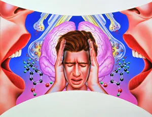
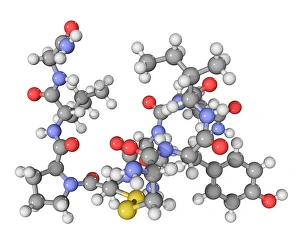
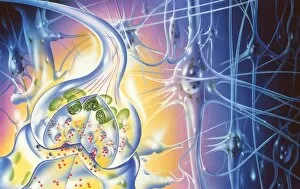
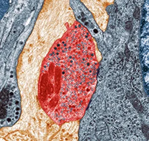
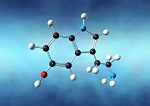
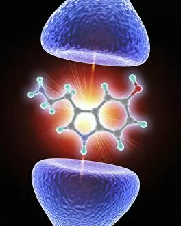
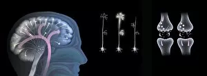
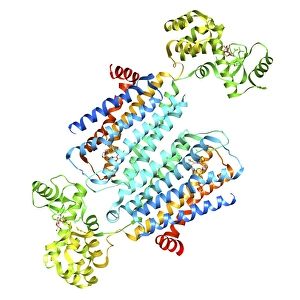
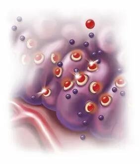
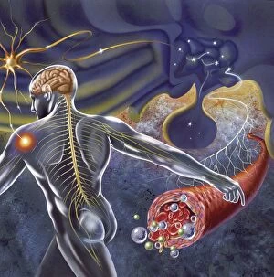
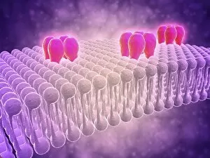
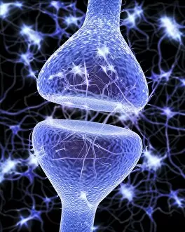
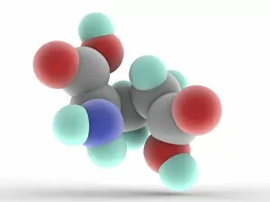
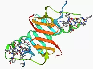
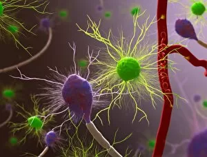
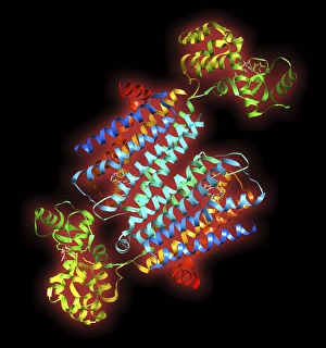
"Exploring the Intricacies of Neurotransmitters: Unveiling the Secrets of Brain Protein Research" The fascinating world of neurotransmitters unravels as scientists delve into brain protein research, seeking to understand the complex mechanisms that govern our thoughts and emotions. Intriguingly, cannabinoid receptor binding emerges as a crucial aspect in this intricate web. Artwork depicting these receptors highlights their role in modulating various physiological processes within our brains. As we journey deeper into the realm of neuroscience, we encounter schizophrenia—a disorder shrouded in mystery. Researchers tirelessly investigate how neurotransmitters contribute to its development and progression, hoping to unlock new treatment avenues for those affected. Amongst these chemical messengers lies oxytocin, known as the "love hormone. " Its unique structure is captured beautifully as a molecule, emphasizing its vital role in social bonding and trust. Nerve synapses become our next stop on this captivating voyage. Through TEM images, we witness the astonishing intricacy with which neurons communicate through electrical impulses and neurotransmitter release—bridging gaps between cells with astonishing precision. Serotonin takes center stage—an influential neurotransmitter responsible for regulating mood and sleep patterns. Visualizing it at a molecular level helps us appreciate its significance in maintaining emotional well-being. Enkephalin crystals shimmer under light micrographs—an opioid peptide offering insights into pain management strategies. Understanding their structures allows researchers to develop novel therapies targeting pain pathways effectively. Delving further into neurochemistry brings us face-to-face with ecstasy's impact on brain function—a topic both intriguing and concerning simultaneously. Scientists strive to comprehend how this recreational drug affects neurotransmission patterns while considering potential long-term consequences. Dopamine receptor D3 C016/4464 captures attention—a key player implicated in reward-motivated behavior and addiction disorders. Studying its structure provides valuable clues towards developing targeted interventions for substance abuse issues plaguing society today.