Fraud Blocker Skip to main content

Nucleus Collection (#17)

"The Nucleus: Unveiling the Mysteries of Life's Command Center" Exploring the intricate world within our cerebellum tissue

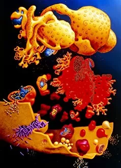
Background imageNucleus Collection: Artwork of ER, golgi body, plasma membrane of cell

Artwork of ER, golgi body, plasma membrane of cell
Illustration of membrane structures in a cell: endoplasmic reticulum, golgi apparatus and plasma membrane. At top, the nucleus is seen with pores, & ribosomes (pink dots) on its surface

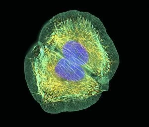
Background imageNucleus Collection: HaCaT culture cell, light micrograph

HaCaT culture cell, light micrograph
HaCaT cell. Immunofluorescence light micrograph of a HaCaT cell dividing into two. The nucleus, which contains the cells genetic information, is purple

Background imageNucleus Collection: Human cell

Human cell, computer-enhanced confocal light micrograph. This unidentified cell has a large nucleus (rounded, centre) and several long projections from its body. Magnification unknown

Background imageNucleus Collection: Cellular clock

Cellular clock. Conceptual artwork of a clock and the internal structures (organelles) of an animal cell. This could represent the rate of metabolic cell reactions

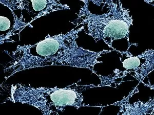
Background imageNucleus Collection: Cells, SEM

Cells, SEM
Cell cytoskeleton. Coloured scanning electron micrograph (SEM) of the cytoskeleton (blue) and nuclei (green) of cultured cells

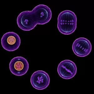
Background imageNucleus Collection: Mitosis, artwork

Mitosis, artwork

Background imageNucleus Collection: Smooth muscle, light micrograph

Smooth muscle, light micrograph
Smooth muscle. Light micrograph of a longitudinal section through smooth muscle from the gut. The cells have typically elongated nuclei. Magnification: x240 when printed at 10 centimetres wide

Background imageNucleus Collection: DNA nucleosomes, molecular model

DNA nucleosomes, molecular model
DNA nucleosomes. Molecular model of four DNA (deoxyribonucleic acid) nucleosomes, known as a tetranucleosome. Nucleosomes are the fundamental repeating units used to package DNA inside cell nuclei

Background imageNucleus Collection: Quantum atom model

Quantum atom model
Conceptual computer artwork of a quantum atom model depicting the sub-atomic particles of quantum physics

Background imageNucleus Collection: Ribosomes, TEM

Ribosomes, TEM
Ribosomes. Coloured transmission electron micrograph (TEM) of ribosomes (blue) passing through pores in a cells nuclear membrane (red)

Background imageNucleus Collection: Fallopian tube, TEM

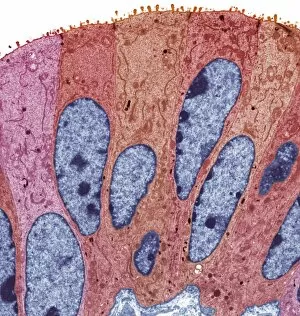
Fallopian tube, TEM
Fallopian tube, coloured transmission electron micrograph (TEM). Section through non-ciliated columnar epithelium from a fallopian tube. The ciliated cells are not shown here

Background imageNucleus Collection: Planetary orbits, artwork

Planetary orbits, artwork
Planetary orbits, conceptual computer artwork

Background imageNucleus Collection: Quantised orbits of the planets

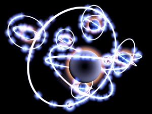
Quantised orbits of the planets
Quantum orbits of the planets. Conceptual computer artwork showing the planets of the solar system on a model of atomic orbitals

Background imageNucleus Collection: Active galaxy, conceptual artwork

Active galaxy, conceptual artwork
Active galaxy, conceptual computer artwork. Active galaxies are typical galaxies (centre), but with a small core of emission in the middle of the galaxy, known as an active galactic nucleus (AGN)

Background imageNucleus Collection: Trachea mucous membrane, SEM

Trachea mucous membrane, SEM
Trachea mucous membrane. Coloured scanning electron micrograph (SEM) of a fractured mucous membrane of the trachea (wind pipe), showing the epithelium and underlying connective tissue

Background imageNucleus Collection: Cerebellum structure, light micrograph

Cerebellum structure, light micrograph
Cerebellum structure. Coloured light micrograph of a section through the highly-folded cerebellum of the brain. The cerebellum comprises three main layers

Background imageNucleus Collection: Mast cell, TEM

Mast cell, TEM
Mast cell, coloured transmission electron micrograph (TEM). Mast cells are a type of white blood cell found in connective tissue

Background imageNucleus Collection: Atomic structure, conceptual artwork

Atomic structure, conceptual artwork
Atomic structure. Conceptual computer artwork of electron orbit paths as rings around the central nucleus (yellow) of an atom

Background imageNucleus Collection: Influenza viruses, TEM

Influenza viruses, TEM
Influenza viruses. Coloured transmission electron micrograph (TEM) of influenza (flu) viruses (yellow) budding from a host cell. The host cell nucleus is black

Background imageNucleus Collection: Lymphocyte white blood cell, SEM

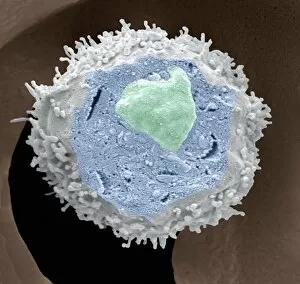
Lymphocyte white blood cell, SEM
Lymphocyte white blood cell. Coloured scanning electron micrograph (SEM) of a section through a lymphocyte, showing the cell nucleus (upper centre) and cytosol (blue)

Background imageNucleus Collection: Comet, historical image

Comet, historical image
Head of the Comet of 1961. The rotating core produced shell-like figures with some bright spots which are larger ice bolders leaving the comet service we know today

Background imageNucleus Collection: Frogspawn, SEM

Frogspawn, SEM
Frogspawn. Coloured scanning electron micrograph (SEM) of a section through frog eggs, showing the large nucleus containing nucleoli, surrounded by yolk

Background imageNucleus Collection: Pancreas tissue, SEM

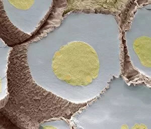
Pancreas tissue, SEM
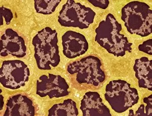
Pancreas tissue. Coloured scanning electron micrograph (SEM) of fractured pancreas tissue, showing numerous acinar cells, containing secretory zymogen granules

Background imageNucleus Collection: Monocyte white blood cells, artwork

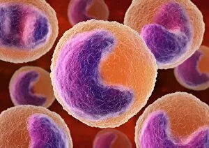
Monocyte white blood cells, artwork
Monocyte white blood cells. Computer artwork of monocyte white blood cells, showing their lobed nuclei (purple, centre). Monocytes, like all white blood cells

Background imageNucleus Collection: Basophil white blood cells, artwork

Basophil white blood cells, artwork
Basophil white blood cells. Computer artwork of basophil white blood cells, showing their lobed nuclei (red). Basophils are the smallest and least common of the white blood cells

Background imageNucleus Collection: Lymph node, TEM

Lymph node, TEM
Lymph node. Coloured transmission electron micrograph (TEM) of a section through the peripheral corona of a lymph nodule, showing numerous small lymphocyte white blood cells (dark)

Background imageNucleus Collection: Cell nucleus, TEM

Cell nucleus, TEM
Cell nucleus. Coloured transmission electron micrograph (TEM) of a section through a cell, showing the nucleus (large, spherical), and mitochondria (green)

Background imageNucleus Collection: Lithium atoms, computer artwork

Lithium atoms, computer artwork
Computer artwork of seven lithium atoms with their nucleus and the three orbiting electrons

Background imageNucleus Collection: Fat cell, TEM

Fat cell, TEM
Fat cell. Coloured Coloured transmission electron micrograph (TEM) of a section through fat (adipose) tissue. Fat cells, or adipocytes

Background imageNucleus Collection: Eosinophil white blood cells, artwork

Eosinophil white blood cells, artwork
Eosinophil white blood cells. Computer artwork of eosinophil white blood cells, showing their lobed nuclei (centre). Eosinophils, like all white blood cells, are part of the immune system

Background imageNucleus Collection: Macrophage white blood cell, TEM

Macrophage white blood cell, TEM
Macrophage white blood cell. Coloured transmission electron micrograph (TEM) of a section through a macrophage from a lymph node, showing the nucleus (red) and numerous lysosomes (black)

Background imageNucleus Collection: Nerve cells, artwork

Nerve cells, artwork
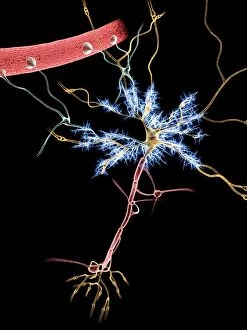
Neuron, nerve cell, firing, computer artwork

Background imageNucleus Collection: Motor neuron, SEM

Motor neuron, SEM
Motor neuron. Scanning electron micrograph (SEM) of section through a motor neuron (nerve cell, dark blue) from the spinal cord, showing its nucleus (light blue, centre) and nucleolus (pink, centre)


All Professionally Made to Order for Quick Shipping

"The Nucleus: Unveiling the Mysteries of Life's Command Center" Exploring the intricate world within our cerebellum tissue, where the nucleus orchestrates every movement and balance. A mesmerizing light micrograph reveals the nucleus at the heart of M51 Whirlpool Galaxy, a cosmic dance guided by its gravitational pull. Witnessing life's beginning through a budding yeast cell, as its nucleus holds the blueprint for growth and reproduction. Delving into the secrets of communication with a nerve cell captured in stunning detail through SEM imaging, showcasing its complex nucleus. Behold the majestic Sombrero Galaxy (Messier 104), where billions of stars revolve around their central nucleus like celestial dancers in perfect harmony. The hippocampus brain tissue unveils memories etched within each neuron's nucleus, shaping who we are and what we remember. Peering into HeLa cells under a light microscope (C017/8299), revealing their remarkable nuclei that have revolutionized medical research. An artistic representation takes us deep into Medulla Oblongata's core—the vital center controlling essential bodily functions—where nuclei reign supreme. Captivating artwork depicts nuclear fission—a powerful force harnessed to generate energy while reminding us of its immense potential and responsibility. Marvel at NGC 4258 spiral galaxy's galactic light show, where vibrant colors illuminate star clusters revolving around their radiant nuclear core. Human cells come alive under scrutiny as their nuclei hold genetic information that shapes our unique characteristics and existence itself. Exploring kidney tubules in section unravels an intricate network governed by countless tiny nuclei working harmoniously to maintain our body’s equilibrium. Intricate yet awe-inspiring, these glimpses into various realms remind us that no matter how vast or minuscule, from galaxies to microscopic cells - all bow to the commanding presence of the nucleus.