Fraud Blocker Skip to main content

Ocelli Collection

Ocelli, the tiny yet remarkable eyes of insects, provide a fascinating glimpse into their world

Background imageOcelli Collection: Honey Bee (Apis mellifera), internal anatomy, cross-section

Honey Bee (Apis mellifera), internal anatomy, cross-section

Background imageOcelli Collection: Bee head, SEM C018 / 0546

Bee head, SEM C018 / 0546
Bee head, coloured scanning electron micrograph (SEM). This is the head of a small bumblebee. Flying insects often have two sets of eyes, coloured pink here

Background imageOcelli Collection: Male bee head C018 / 3568

Male bee head C018 / 3568
Male bee head. Close-up photograph of a male bee head (Megachile lanata). This specimen was found in Hawaii. The species was originally from Africa and has since spread to the Caribbean

Background imageOcelli Collection: Illustration, Butterfly with eyespots on wings

Illustration, Butterfly with eyespots on wings

Background imageOcelli Collection: Mercury with the head of Argus, Hieronymus Cock, Matthys Cock, c. 1551 - before 1558

Mercury with the head of Argus, Hieronymus Cock, Matthys Cock, c. 1551 - before 1558

Background imageOcelli Collection: Female bumblebee, Bombus auricomas C018 / 3579

Female bumblebee, Bombus auricomas C018 / 3579
Female bumblebee, Bombus auricomas. This species is native to North America and inhabits urban areas. Photographed by the USGS Bee Inventory and Monitoring Lab

Background imageOcelli Collection: Female sweat bee head with pollen C018 / 3573

Female sweat bee head with pollen C018 / 3573
Female sweat bee head with pollen. Close-up photograph of a female sweat bee (Halictus ligatus) covered in pollen. This specimen is native to North America and was found in Pennsylvania

Background imageOcelli Collection: Parasitic fly, SEM Z340 / 0605

Parasitic fly, SEM Z340 / 0605
Parasitic fly. Coloured scanning electron micrograph (SEM) of the fly Ornithomyia avicularia. This is a parasite of nesting birds, especially house martins and swallows

Background imageOcelli Collection: Spider simple eyes, SEM C018 / 0560

Spider simple eyes, SEM C018 / 0560
Spider simple eyes, coloured scanning electron micrograph (SEM). The arrangement of eyes differs widely between spiders, but in general they have eight eyes (here)

Background imageOcelli Collection: Wasp simple eyes, SEM C018 / 0559

Wasp simple eyes, SEM C018 / 0559
Wasp simple eyes, coloured scanning electron micrograph (SEM). These three dorsal ocelli ( simple eyes ) are located on top of the wasps head

Background imageOcelli Collection: Male bee C018 / 3571

Male bee C018 / 3571
Male bee. Close-up photograph of a male bee head (Anthophora affabilis). This specimen is native to the US and was found in Badlands National Park, South Dakota

Background imageOcelli Collection: Female bee head C018 / 3570

Female bee head C018 / 3570
Female bee head. Close-up photograph of a female bee head (Megachile lanata). This specimen was found in Cuba. The species was originally from Africa and has since spread to the Caribbean

Background imageOcelli Collection: Stick mantis, Borneo C013 / 4982

Stick mantis, Borneo C013 / 4982
Stick mantis (Paratoxodera sp.) This mantis resembles a plant twig. It remains concealed while waiting for prey to move within its reach

Background imageOcelli Collection: Collembola ocelli, springtail

Collembola ocelli, springtail
Scanning electron microscope image of the springtail with simple eyes (x 1.2K)

Background imageOcelli Collection: Jumping spiders eye, SEM

Jumping spiders eye, SEM
Jumping spiders eye. Coloured scanning electron micrograph (SEM) of the simple eye (ocelli, orange) of a jumping spider (family Salticidae)

Background imageOcelli Collection: Jumping spiders eyes, SEM

Jumping spiders eyes, SEM
Jumping spiders eyes. Coloured scanning electron micrograph (SEM) of the eyes (brown) of a jumping spider (family Salticidae)

Background imageOcelli Collection: Soldier ant jaws, SEM

Soldier ant jaws, SEM
Soldier ant jaws. Coloured scanning electron micrograph (SEM) of the head of a leaf-cutter ant (Atta sexdens). This is a soldier ant

Background imageOcelli Collection: Caterpillar head, SEM

Caterpillar head, SEM
Caterpillar head. Coloured scanning electron micrograph (SEM) of the head of a caterpillar. Caterpillars (order Lepidoptera) are the larval stage of the life cycle of a butterfly or moth

Background imageOcelli Collection: Fly eyes, SEM

Fly eyes, SEM
Fly eyes. Coloured scanning electron micrograph (SEM) of the simple eyes (ocelli) of a hover fly (family Syrphidae). There are three

Background imageOcelli Collection: Adult fruit fly hatching, SEM

Adult fruit fly hatching, SEM
Fruit fly hatching. Coloured scanning electron micrograph (SEM) of an imago (adult) of the fruit fly Drosophila melanogaster (wild type, Oregon R), at eclosion (hatching)

Background imageOcelli Collection: Argus Pheasant feather

Argus Pheasant feather
Argus secondary wing feathers showing ocelli. See also Fig 56. Page 143 Vol II of Charles Darwin " The Descent of Man and Selection in Relation to Sex" 1871

Background imageOcelli Collection: Peacock Pavo cristatus displaying tail

Peacock Pavo cristatus displaying tail
Male peacock, Pavo cristatus, displaying his ocellated tail feathers. The peacocks tail, cumbersome and expensive to produce, required an explanation under Darwins theory of natural selection

Background imageOcelli Collection: Attacus atlas moth incipient mimicry grn

Attacus atlas moth incipient mimicry grn
Wing tip of an atlas moth (Attacus atlas) of Northern India showing the incipient ocelli and markings which appear to resemble a snakes head

Background imageOcelli Collection: Argus feather and Darwin illustration

Argus feather and Darwin illustration
Composite photograph of Argus secondary wing feather left and right Fig 56. Page 143 Vol II of Charles Darwin " The Descent of Man and Selection in Relation to Sex" 1871

Background imageOcelli Collection: 1797 Atlas Moth illustration

1797 Atlas Moth illustration
Copperplate print with contemporary hand colouring from Betruchs " Bilderbuch fur kinder". It shows the worlds largest moth (wing area), the Atlas moth


All Professionally Made to Order for Quick Shipping

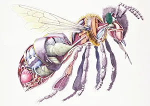
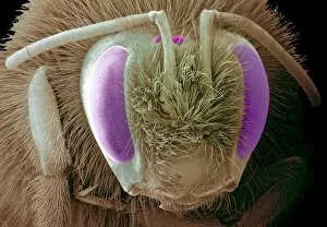
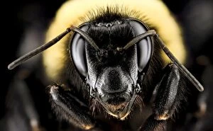
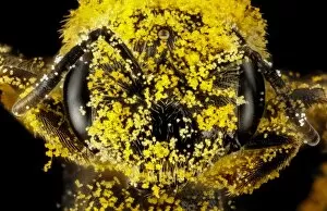
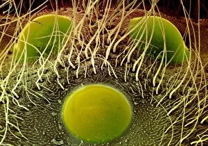
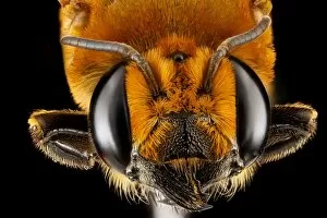
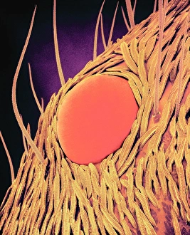
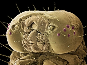
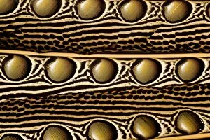
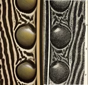
Ocelli, the tiny yet remarkable eyes of insects, provide a fascinating glimpse into their world. Found in various species such as the Honey Bee (Apis mellifera), these specialized organs play a crucial role in their survival. When we delve into the internal anatomy of these creatures and examine cross-sections of their heads under scanning electron microscopes, we are astounded by the intricate details. The SEM images reveal the complex structure on a male bee's head (C018 / 3568) and even offer a closer look at those present on female bees (C018 / 3570). Butterflies also possess ocelli, which can be seen as captivating eyespots adorning their delicate wings. These illustrations remind us of nature's artistry and how it has shaped these beautiful creatures throughout evolution. Interestingly, ancient mythology intertwines with our understanding of ocelli. In one depiction from c. 1551 - before 1558 by Hieronymus Cock and Matthys Cock, Mercury is portrayed with the head of Argus – highlighting both human fascination and imagination surrounding these unique visual organs. Moving beyond bees and butterflies, other insects like bumblebees (Bombus auricomas C018 / 3579) and sweat bees also exhibit distinct ocellar structures that aid them in navigation or detecting pollen-covered surfaces (C018 / 3573). Even parasitic flies have evolved to utilize ocelli for their own purposes (SEM Z340 / 0605). Not limited to just insects, spiders too possess simple eyes known as ocelli that allow them to perceive light variations in their surroundings (SEM C018 /0560). Similarly, wasps rely on this simplified vision system for basic orientation tasks (SEM C018/0559). As we explore further into this realm of insect vision systems through microscopic lenses or artistic interpretations alike; each image reveals the intricate beauty and complexity of ocelli.