Fraud Blocker Skip to main content

Oesophagus Collection

"The Oesophagus: A Journey Through Various Anatomy Artworks" Bee anatomy

Background imageOesophagus Collection: Bee anatomy, artwork

Bee anatomy, artwork
Bee anatomy. Computer artwork showing the internal anatomy of a honeybee (Apis mellifera). Nerve fibres are blue. The wings are attached to the thorax

Background imageOesophagus Collection: Deer anatomy, artwork

Deer anatomy, artwork
Deer anatomy. Computer artwork showing the internal anatomy of a deer (family Cervidae). Anatomical features shown here include the skeleton, lungs, intestines, bladder

Background imageOesophagus Collection: Human digestive system, artwork

Human digestive system, artwork. At top, food from the mouth forms a bolus that is swallowed down the oesophagus. At centre, the bolus is about to enter the stomach (arrow)

Background imageOesophagus Collection: Col X-ray of object (safety pin) lodged in throat

Col X-ray of object (safety pin) lodged in throat
Obstruction in throat. Coloured X-ray of an object (safety pin) lodged in the oesophagus of a woman. In this side view of the neck, the swallowed object is seen at centre right

Background imageOesophagus Collection: Fig.1.2. Stinging Cubomedusae; F.3.4. Its Long Tentacles; F. 5. Sea Medusae with... 1813

Fig.1.2. Stinging Cubomedusae; F.3.4. Its Long Tentacles; F. 5. Sea Medusae with... 1813
Fig.1.2. Stinging Cubomedusae; F.3.4. Its Long Tentacles; F. 5. Sea Medusae with Long Tentacles; F.6. Curly Fringe of a Large Brazilian Cubomedusae; F.7.Warty Sea Medusae; F.8

Background imageOesophagus Collection: F.1. Japanese 'Golovach'; F.2. Its Head; F.3. From Below, Where Small Nipples on... 1813

F.1. Japanese "Golovach"; F.2. Its Head; F.3. From Below, Where Small Nipples on... 1813
F.1. Japanese "Golovach"; F.2. Its Head; F.3. From Below, Where Small Nipples on Its Gullet Are Visible, 1813. In 1803-06, Captain I. F

Background imageOesophagus Collection: Upper respiratory tract, artwork

Upper respiratory tract, artwork
Upper respiratory tract, computer artwork

Background imageOesophagus Collection: Two Views of the Head, 1746. Creator: Jacques Fabien Gautier Dagoty

Two Views of the Head, 1746. Creator: Jacques Fabien Gautier Dagoty
Two Views of the Head, 1746. Plate 4 in Myologie Complette en Couleur et Grandeur Naturelle, a work on myology, (study of the muscular system)

Background imageOesophagus Collection: Plate from Atlas and Epitome of Operative Surgery by Dr

Plate from Atlas and Epitome of Operative Surgery by Dr
CHT235342 Plate from Atlas and Epitome of Operative Surgery by Dr. Otto Zuckerkandl (1861-1921) 1910 (coloured engraving) by French School, (20th century); Bibliotheque de la Faculte de Medecine

Background imageOesophagus Collection: Diagram of the abdominal cavity in the human body with the liver removed

Diagram of the abdominal cavity in the human body with the liver removed

Background imageOesophagus Collection: Diagram showing human liver, stomach, gallbladder and pancreas

Diagram showing human liver, stomach, gallbladder and pancreas

Background imageOesophagus Collection: Human organs, artwork

Human organs, artwork
Human organs, computer artwork

Background imageOesophagus Collection: Human neck anatomy, artwork C017 / 7259

Human neck anatomy, artwork C017 / 7259
Human neck anatomy, artwork. This dissection of the neck shows the larynx (voicebox) at the upper end of the trachea (windpipe, blue)

Background imageOesophagus Collection: Upper respiratory tract, artwork

Upper respiratory tract, artwork
Upper respiratory tract, computer artwork

Background imageOesophagus Collection: Spider anatomy, artwork

Spider anatomy, artwork
Spider anatomy. Computer artwork showing the main organs of a typical female spider

Background imageOesophagus Collection: Larynx anatomy, artwork

Larynx anatomy, artwork
Larynx anatomy. Artwork of the anatomy of the human larynx (voicebox) showing the production of vocal sounds (yellow arrows)

Background imageOesophagus Collection: Cow anatomy, artwork

Cow anatomy, artwork
Cow anatomy. Computer artwork showing the internal anatomy of a domesticated cow (Bos primigenius). Anatomical features shown here include the skeleton, lungs, intestines, bladder

Background imageOesophagus Collection: Cross section of human stomach

Cross section of human stomach

Background imageOesophagus Collection: Cross section illustration of internal anatomy of female lizard

Cross section illustration of internal anatomy of female lizard

Background imageOesophagus Collection: Cross section illustration of mouse internal organs

Cross section illustration of mouse internal organs

Background imageOesophagus Collection: Digital illustration of woman showing brain, oesophagus, heart, stomach, intestine, veins and muscle

Digital illustration of woman showing brain, oesophagus, heart, stomach, intestine, veins and muscle

Background imageOesophagus Collection: Illustration of contractions of muscles in the wall of the oesophagus (peristalsis)

Illustration of contractions of muscles in the wall of the oesophagus (peristalsis)

Background imageOesophagus Collection: Human respiratory system

Human respiratory system

Background imageOesophagus Collection: Picture No. 10890916

Picture No. 10890916
Egg Eating Snake - the egg has now been swallowed and the snake arches its neck to crush it in the oesophagus (Dasypeltis fasciata) Date:

Background imageOesophagus Collection: Human anatomy, artwork F007 / 3985

Human anatomy, artwork F007 / 3985
Human anatomy, computer artwork

Background imageOesophagus Collection: Female anatomy, artwork F007 / 3678

Female anatomy, artwork F007 / 3678
Female anatomy, computer artwork

Background imageOesophagus Collection: Human anatomy, artwork F007 / 3785

Human anatomy, artwork F007 / 3785
Human anatomy, computer artwork

Background imageOesophagus Collection: Human anatomy, artwork F007 / 4089

Human anatomy, artwork F007 / 4089
Human anatomy, computer artwork

Background imageOesophagus Collection: Female anatomy, artwork F007 / 3492

Female anatomy, artwork F007 / 3492
Female anatomy, computer artwork

Background imageOesophagus Collection: Healthy digestive system, artwork F006 / 3460

Healthy digestive system, artwork F006 / 3460
Healthy digestive system, computer artwork

Background imageOesophagus Collection: Small intestine and stomach, artwork F007 / 7235

Small intestine and stomach, artwork F007 / 7235
Small intestine and stomach, computer artwork

Background imageOesophagus Collection: Abdominal pain, artwork F007 / 7236

Abdominal pain, artwork F007 / 7236
Abdominal pain, computer artwork

Background imageOesophagus Collection: Female anatomy, artwork F007 / 4404

Female anatomy, artwork F007 / 4404
Female anatomy, computer artwork

Background imageOesophagus Collection: Man drinking, artwork F007 / 6105

Man drinking, artwork F007 / 6105
Man drinking, computer artwork

Background imageOesophagus Collection: Man drinking, artwork F007 / 6107

Man drinking, artwork F007 / 6107
Man drinking, computer artwork

Background imageOesophagus Collection: Man drinking, artwork F007 / 6106

Man drinking, artwork F007 / 6106
Man drinking, computer artwork

Background imageOesophagus Collection: Man drinking, artwork F007 / 6112

Man drinking, artwork F007 / 6112
Man drinking, computer artwork

Background imageOesophagus Collection: Man drinking, artwork F007 / 6109

Man drinking, artwork F007 / 6109
Man drinking, computer artwork

Background imageOesophagus Collection: Man drinking, artwork F007 / 6116

Man drinking, artwork F007 / 6116
Man drinking, computer artwork

Background imageOesophagus Collection: Man drinking, artwork F007 / 6111

Man drinking, artwork F007 / 6111
Man drinking, computer artwork

Background imageOesophagus Collection: Man drinking, artwork F007 / 6113

Man drinking, artwork F007 / 6113
Man drinking, computer artwork

Background imageOesophagus Collection: Man drinking, artwork F007 / 6114

Man drinking, artwork F007 / 6114
Man drinking, computer artwork

Background imageOesophagus Collection: Female anatomy, artwork F007 / 4502

Female anatomy, artwork F007 / 4502
Female anatomy, computer artwork

Background imageOesophagus Collection: Man drinking, artwork F007 / 6108

Man drinking, artwork F007 / 6108
Man drinking, computer artwork

Background imageOesophagus Collection: Man drinking, artwork F007 / 6110

Man drinking, artwork F007 / 6110
Man drinking, computer artwork

Background imageOesophagus Collection: Female anatomy, artwork F007 / 5502

Female anatomy, artwork F007 / 5502
Female anatomy, computer artwork

Background imageOesophagus Collection: Male anatomy, artwork F008 / 1394

Male anatomy, artwork F008 / 1394
Male anatomy, computer artwork

Background imageOesophagus Collection: Male anatomy, artwork F008 / 1393

Male anatomy, artwork F008 / 1393
Male anatomy, computer artwork


All Professionally Made to Order for Quick Shipping

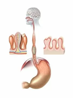
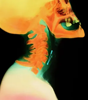
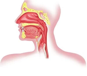
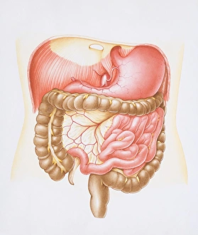
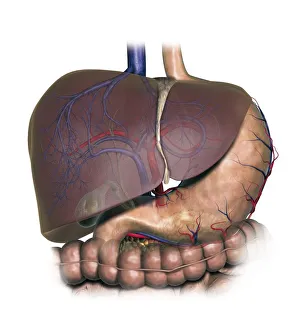
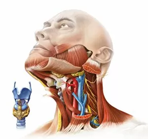
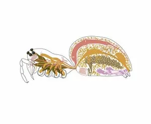
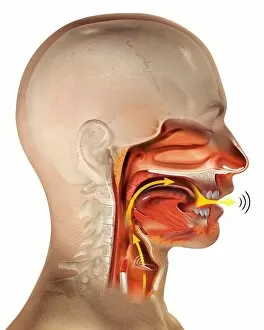
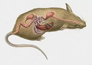
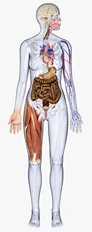
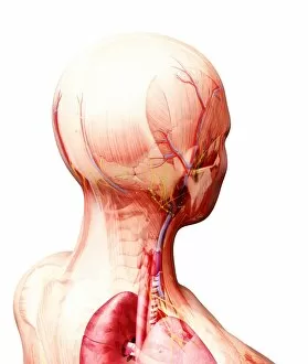
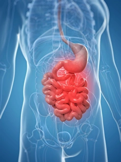
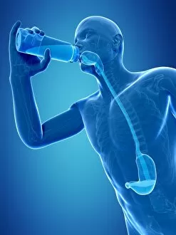
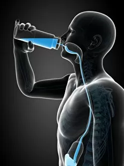
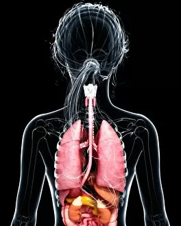
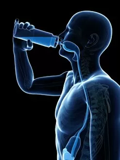
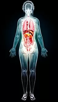
"The Oesophagus: A Journey Through Various Anatomy Artworks" Bee anatomy, artwork: Delve into the intricate world of bees and discover how their oesophagus plays a vital role in their survival. Human digestive system, artwork: Explore the fascinating intricacies of the human digestive system, with a focus on the oesophagus as it transports food from mouth to stomach. Col X-ray of object (safety pin) lodged in throat: Witness a captivating X-ray image revealing an unexpected obstacle - a safety pin lodged in the oesophagus - highlighting its vulnerability and importance. Dog anatomy, artwork: Discover how man's best friend also possesses an oesophagus that aids in digestion and nourishment for our beloved canines. Two Views of the Head, 1746 by Jacques Fabien Gautier Dagoty: Step back in time with this stunning artwork showcasing detailed depictions of various anatomical structures including the oesophagus within the context of human physiology. Plate from Atlas and Epitome of Operative Surgery by Dr. . : Uncover surgical insights through this illustrative plate depicting operative procedures involving the oesophagus; witness medical advancements at work. Diagram showing human liver, stomach, gallbladder and pancreas: Understand how these essential organs interact with each other while being connected to one another via the crucial pathway provided by our trusty oesophagus. Human neck anatomy, artwork C017 / 7259: Marvel at artistic renderings capturing every detail of our neck's complex structure where we find not only blood vessels but also our resilient esophageal tube guiding sustenance down towards nourishment or potential danger if obstructed. Deer anatomy, artwork: Embark on a journey through nature's wonders as we explore deer anatomy including their unique adaptation when it comes to the oesophagus, enabling them to thrive in their environments.