Fraud Blocker Skip to main content

Oncological Collection (#4)

"Unveiling the Hidden Battles: A Glimpse into the World of Oncology" Acute promyelocytic leukaemia, micrograph: Witnessing the intricate battle within

Background imageOncological Collection: Mouth cancer cell, SEM

Mouth cancer cell, SEM
Mouth cancer cell. Coloured scanning electron micrograph (SEM) showing the surface of a squamous cell carcinoma (cancer) cell from a human mouth

Background imageOncological Collection: Liver cancer cell, SEM

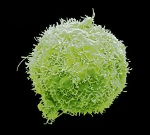
Liver cancer cell, SEM
Liver cancer cell. Coloured scanning electron micrograph (SEM) of a hepatocellular carcinoma (HCC) cell, showing the numerous filopodia (hair-like) covering its surface

Background imageOncological Collection: Dividing liver cancer cell, SEM

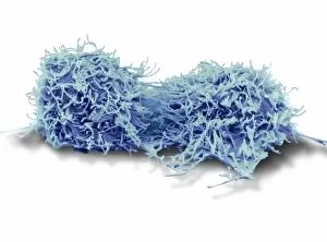
Dividing liver cancer cell, SEM
Dividing liver cancer cell. Coloured scanning electron micrograph (SEM) of a hepatocellular carcinoma (HCC) cell undergoing mitosis (nuclear division)

Background imageOncological Collection: Secondary bowel cancer, endoscopic view C016 / 8337

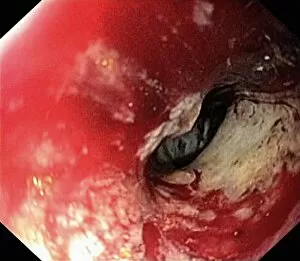
Secondary bowel cancer, endoscopic view C016 / 8337
Secondary bowel cancer. Endoscopic view of the interior of a patients descending duodenum (first part of the small intestine), showing cancer that has spread (metastasised) from the pancreas

Background imageOncological Collection: Colon cancer, endoscopic view C016 / 8307

Colon cancer, endoscopic view C016 / 8307
Colon cancer. Endoscopic view of a malignant (cancerous) adenocarcinoma (lower right) in a patients ascending colon (large intestine)

Background imageOncological Collection: Radiotherapy, conceptual image C013 / 5587

Radiotherapy, conceptual image C013 / 5587
Radiotherapy, conceptual image. Computer artwork representing the use of ionising radiation to kill cancerous cells (centre)

Background imageOncological Collection: Dividing cancer cell, SEM C014 / 0361

Dividing cancer cell, SEM C014 / 0361
Dividing cancer cell. Coloured scanning electron micrograph (SEM) of a colorectal cancer cell undergoing mitosis (nuclear division) and splitting into two daughter cells (left and right)

Background imageOncological Collection: Dividing brain cancer cells, SEM C014 / 0354

Dividing brain cancer cells, SEM C014 / 0354
Dividing brain cancer cells. Coloured scanning electron micrograph (SEM) of a cancerous astrocyte brain cell that has just undergone cytokinesis (cell division)

Background imageOncological Collection: Dividing bone cancer cell, SEM

Dividing bone cancer cell, SEM
SEM Dividing Osteosarcoma cells Osteosarcoma is an aggressive malignant neoplasm arising from primitive transformed cells of mesenchymal origin (and thus a sarcoma)

Background imageOncological Collection: Cisplatin drug molecule

Cisplatin drug molecule. Computer artwork showing the structure of a molecule of the chemotherapy drug Cisplatin. This drug is used to treat a number of cancers, including sarcomas

Background imageOncological Collection: Cancer cell, SEM C014 / 0628

Cancer cell, SEM C014 / 0628
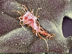
Cancer cell. Coloured scanning electron micrograph (SEM) of a carcinoma cancer cell (pink) migrating (metastasising) along a layer of normal epithelial cells (purple)

Background imageOncological Collection: Cancer cell, SEM C014 / 0627

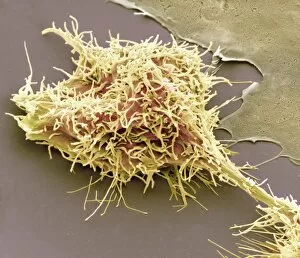
Cancer cell, SEM C014 / 0627
Cancer cell. Coloured scanning electron micrograph (SEM) of a carcinoma cancer cell (yellow) migrating (metastasising) along a layer of normal epithelial cells (brown)

Background imageOncological Collection: Throat cancer, artwork C013 / 5939

Throat cancer, artwork C013 / 5939
Throat cancer. Cutaway computer artwork of a persons head, showing cancer (yellow) in the pharynx (centre) and oesophagus (lower centre)

Background imageOncological Collection: Bone cancer cell, SEM

Bone cancer cell, SEM
Bone cancer cell. Coloured scanning electron micrograph of a fibrosarcoma (fibroblastic sarcoma) cell, showing the numerous filopodia (hair-like) covering its surface

Background imageOncological Collection: Intestinal tumour, artwork C015 / 6451

Intestinal tumour, artwork C015 / 6451
Intestinal tumour. Computer artwork of a tumour on the internal wall of an intestine

Background imageOncological Collection: Chronic myeloid leukaemia, micrograph C015 / 6227

Chronic myeloid leukaemia, micrograph C015 / 6227
Chronic myeloid leukaemia. Light micrograph of bone marrow from a patient with chronic myeloid leukaemia (CML). This cancer affects the myeloid tissue (bone marrow)

Background imageOncological Collection: Acute myeloid leukaemia C015 / 6225

Acute myeloid leukaemia C015 / 6225
Acute myeloid leukaemia. Light micrograph of bone marrow from a patient with acute myeloid leukaemia (AML). This cancer affects the myeloid tissue (bone marrow)

Background imageOncological Collection: Breast cancer protein molecule C016 / 4455

Breast cancer protein molecule C016 / 4455
Breast cancer protein. Molecular model of breast cancer type 1 susceptibility (BRCA1) protein. This protein is found in breast and other tissue

Background imageOncological Collection: Breast cancer protein molecule C016 / 4454

Breast cancer protein molecule C016 / 4454
Breast cancer protein. Molecular model of breast cancer type 1 susceptibility (BRCA1) protein. This protein is found in breast and other tissue

Background imageOncological Collection: Breast cancer protein molecule C016 / 4453

Breast cancer protein molecule C016 / 4453
Breast cancer protein. Molecular model of breast cancer type 1 susceptibility (BRCA1) protein. This protein is found in breast and other tissue

Background imageOncological Collection: Breast cancer protein molecule C016 / 4450

Breast cancer protein molecule C016 / 4450
Breast cancer protein. Molecular model of breast cancer type 1 susceptibility (BRCA1) protein. This protein is found in breast and other tissue

Background imageOncological Collection: Brain cancer, MRI scan C016 / 4438

Brain cancer, MRI scan C016 / 4438
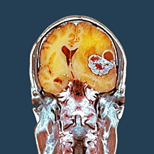
Brain cancer. Coloured coronal magnetic resonance imaging (MRI) scan through the head of a 48 year old male patient with a glioblastoma (red)

Background imageOncological Collection: Brain cancer, MRI scan C016 / 4436

Brain cancer, MRI scan C016 / 4436
Brain cancer. Coloured coronal magnetic resonance imaging (MRI) scan through the head of a 48 year old male patient with a glioblastoma (centre right)

Background imageOncological Collection: Brain cancer, MRI scan C016 / 4437

Brain cancer, MRI scan C016 / 4437
Brain cancer. Coloured coronal magnetic resonance imaging (MRI) scan through the head of a 48 year old male patient with a glioblastoma (orange)

Background imageOncological Collection: DNA and tumour suppressor complex C014 / 0014

DNA and tumour suppressor complex C014 / 0014
DNA and tumour suppressor complex. Molecular model showing a molecule of the p53 tumour suppressor protein (green, centre) bound to a damaged DNA (deoxyribonucleic acid) strand

Background imageOncological Collection: Bexarotene chemotherapy drug molecule C013 / 7785

Bexarotene chemotherapy drug molecule C013 / 7785
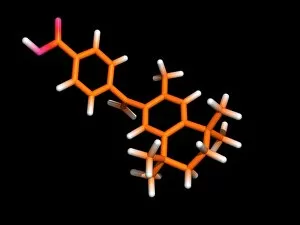
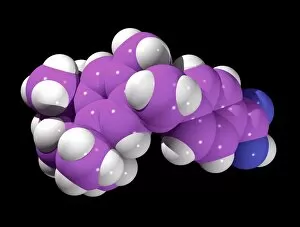
Bexarotene chemotherapy drug molecule. Computer model showing the structure of a molecule of the antineoplastic drug bexarotene (C24H28O2)

Background imageOncological Collection: Bexarotene chemotherapy drug molecule C013 / 7784

Bexarotene chemotherapy drug molecule C013 / 7784
Bexarotene chemotherapy drug molecule. Computer model showing the structure of a molecule of the antineoplastic drug bexarotene (C24H28O2)

Background imageOncological Collection: Radiotherapy treatment C014 / 0164

Radiotherapy treatment C014 / 0164
Radiotherapy treatment. Cancer patient positioned under a linear accelerator in a radiotherapy suite. Linear accelerators are used to produce radiation to treat cancer

Background imageOncological Collection: Lung cancer cell division, artwork C013 / 4630

Lung cancer cell division, artwork C013 / 4630
Lung cancer cell division. Computer artwork of a lung cancer cell during cell division (cytokinesis). The two daughter cells remain temporarily joined by a cytoplasmic bridge (centre)

Background imageOncological Collection: Cancer cell, artwork C013 / 4622

Cancer cell, artwork C013 / 4622
Cancer cell. Computer artwork of a single typical cancer cell in the human body. The uneven surface and cytoplasmic projections are characteristic of cancer cells

Background imageOncological Collection: Bone marrow transplant, conceptual image

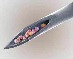
Bone marrow transplant, conceptual image. Composite coloured scanning electron micrograph (SEM) of a needle with bone marrow cells inside

Background imageOncological Collection: Large tumour of the head, 18th century

Large tumour of the head, 18th century


All Professionally Made to Order for Quick Shipping

"Unveiling the Hidden Battles: A Glimpse into the World of Oncology" Acute promyelocytic leukaemia, micrograph: Witnessing the intricate battle within, as microscopic warriors fight against acute promyelocytic leukaemia. Brain tumour, 3D-MRI scan: Peering into the depths of complexity, a three-dimensional MRI scan reveals the enigmatic presence of a brain tumour. Breast Cancer. Oil Painting C017 / 1273: Transcending science through artistry, an oil painting captures the essence and resilience in the face of breast cancer. Brain cancer, DTI and 3D CT scans C016 / 6414: Merging technology's prowess with medical expertise, DTI and 3D CT scans expose the intricate web woven by brain cancer. Cardiac lymphoma, MRI scans: Unmasking an elusive foe that lurks within our hearts - cardiac lymphoma unraveled through revealing MRI scans. Throat cancer, X-rays: Illuminating shadows cast upon vocal cords; X-rays shed light on throat cancer's silent invasion. Vaginal cancer cells SEM C014 /0374-0378): Journeying to unseen realms where life hangs in balance - scanning electron microscopy unveils vaginal cancer cells' haunting beauty and danger simultaneously. Dividing liver cancer cell SEM : Witnessing cellular division amidst chaos – scanning electron microscopy exposes dividing liver cancer cells' relentless propagation. Colorectal Cancer Cell SEM : Exploring nature's hidden battlegrounds – scanning electron microscopy uncovers colorectal cancer cells' formidable presence up close. In this captivating journey through oncological landscapes captured by various imaging techniques like micrographs, MRI scans or SEMs we get a glimpse into battles fought at microscopic levels against diseases like acute promyelocytic leukemia or brain tumors.