Fraud Blocker Skip to main content

Oncology Collection (page 8)

Oncology: Unveiling the Battle Within In the realm of oncology, a fierce war is waged between T lymphocytes and cancer cells

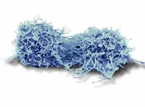
Background imageOncology Collection: Dividing liver cancer cell, SEM

Dividing liver cancer cell, SEM
Dividing liver cancer cell. Coloured scanning electron micrograph (SEM) of a hepatocellular carcinoma (HCC) cell undergoing mitosis (nuclear division)

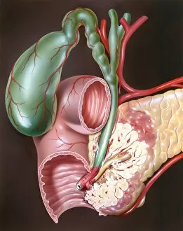
Background imageOncology Collection: Pancreatic cancer, artwork C016 / 8735

Pancreatic cancer, artwork C016 / 8735
Pancreatic cancer. Artwork of part of the digestive system, showing a carcinoma (white, lower right) of the head of the pancreas (light brown)

Background imageOncology Collection: Gastrointestinal disorders, artwork C016 / 8734

Gastrointestinal disorders, artwork C016 / 8734
Gastrointestinal disorders. Artwork of part of the digestive system, showing ulcers on the internal lining of the stomach (centre right) and the internal lining of the duodenum (centre left)

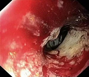
Background imageOncology Collection: Secondary bowel cancer, endoscopic view C016 / 8337

Secondary bowel cancer, endoscopic view C016 / 8337
Secondary bowel cancer. Endoscopic view of the interior of a patients descending duodenum (first part of the small intestine), showing cancer that has spread (metastasised) from the pancreas

Background imageOncology Collection: Colon cancer, endoscopic view C016 / 8307

Colon cancer, endoscopic view C016 / 8307
Colon cancer. Endoscopic view of a malignant (cancerous) adenocarcinoma (lower right) in a patients ascending colon (large intestine)

Background imageOncology Collection: Blood vessel formation, artwork

Blood vessel formation, artwork
Blood vessel formation. Artwork showing malignant (cancerous) tumour cells promoting the formation of new blood vessels, a process known as angiogenesis

Background imageOncology Collection: Radiotherapy, conceptual image C013 / 5587

Radiotherapy, conceptual image C013 / 5587
Radiotherapy, conceptual image. Computer artwork representing the use of ionising radiation to kill cancerous cells (centre)

Background imageOncology Collection: Dividing cancer cell, SEM C014 / 0361

Dividing cancer cell, SEM C014 / 0361
Dividing cancer cell. Coloured scanning electron micrograph (SEM) of a colorectal cancer cell undergoing mitosis (nuclear division) and splitting into two daughter cells (left and right)

Background imageOncology Collection: Dividing brain cancer cells, SEM C014 / 0354

Dividing brain cancer cells, SEM C014 / 0354
Dividing brain cancer cells. Coloured scanning electron micrograph (SEM) of a cancerous astrocyte brain cell that has just undergone cytokinesis (cell division)

Background imageOncology Collection: Dividing bone cancer cell, SEM

Dividing bone cancer cell, SEM
SEM Dividing Osteosarcoma cells Osteosarcoma is an aggressive malignant neoplasm arising from primitive transformed cells of mesenchymal origin (and thus a sarcoma)

Background imageOncology Collection: Cisplatin drug molecule

Cisplatin drug molecule. Computer artwork showing the structure of a molecule of the chemotherapy drug Cisplatin. This drug is used to treat a number of cancers, including sarcomas

Background imageOncology Collection: Cervical cancer, light micrograph C015 / 7000

Cervical cancer, light micrograph C015 / 7000
Cervical cancer. Light micrograph of a section through tissue from the cervix (neck of the womb) showing intraepithelial carcinoma (centre), a form of cancer

Background imageOncology Collection: Cervical cancer, light micrograph C015 / 6999

Cervical cancer, light micrograph C015 / 6999
Cervical cancer. Light micrograph of a section through tissue from the cervix (neck of the womb) showing intraepithelial carcinoma (across centre), a form of cancer

Background imageOncology Collection: Cervical cancer, light micrograph C015 / 6998

Cervical cancer, light micrograph C015 / 6998
Cervical cancer. Light micrograph of a section through tissue from the cervix (neck of the womb) showing intraepithelial carcinoma (dark areas), a form of cancer

Background imageOncology Collection: Cervical cancer, light micrograph C015 / 6997

Cervical cancer, light micrograph C015 / 6997
Cervical cancer. Light micrograph of a section through tissue from the cervix (neck of the womb) showing intraepithelial carcinoma (across centre), a form of cancer

Background imageOncology Collection: Methyltransferase complexed with DNA C016 / 2033

Methyltransferase complexed with DNA C016 / 2033
Methyltransferase complexed with DNA, molecular model. The strand of DNA (deoxyribonucleic acid, green and blue) is enclosed by DNA methyltransferase 1 (DNMT-1, green and pink)

Background imageOncology Collection: Newcastle disease virus protein C015 / 6912

Newcastle disease virus protein C015 / 6912
Newcastle disease virus protein, molecular model. This is a hemagglutinin-neuromidase (HN) protein that enables the virus to bind to and infect host cells

Background imageOncology Collection: Cancer of the chorion C015 / 6905

Cancer of the chorion C015 / 6905
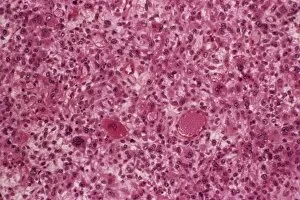
Cancer of the chorion. Light micrograph of a section through a sarcoma of the chorion, a malignant (cancerous) tumour of the membrane (chorion) that surrounds a developing embryo

Background imageOncology Collection: Methyltransferase complexed with DNA C016 / 2032

Methyltransferase complexed with DNA C016 / 2032
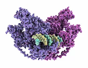
Methyltransferase complexed with DNA, molecular model. The strand of DNA (deoxyribonucleic acid, green and yellow) is enclosed by DNA methyltransferase 1 (DNMT-1, purple and pink)

Background imageOncology Collection: Cancer research

Cancer research
MODEL RELEASED. Cancer research. Researcher standing in front of an immunoassay machine. An immunoassay (antigen-antibody assay) uses antibodies to detect the presence of a certain protein in a sample

Background imageOncology Collection: Cervical smear showing endometrial cancer C015 / 6750

Cervical smear showing endometrial cancer C015 / 6750
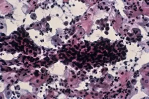
Cervical smear showing endometrial cancer. Light micrograph of cells from a cervical smear test showing endometrial sarcoma. This is a cancer of the lining of the womb, the endometrium

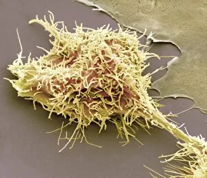
Background imageOncology Collection: Cancer cell, SEM C014 / 0628

Cancer cell, SEM C014 / 0628
Cancer cell. Coloured scanning electron micrograph (SEM) of a carcinoma cancer cell (pink) migrating (metastasising) along a layer of normal epithelial cells (purple)

Background imageOncology Collection: Cancer cell, SEM C014 / 0627

Cancer cell, SEM C014 / 0627
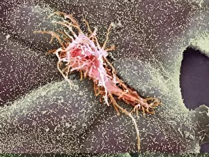
Cancer cell. Coloured scanning electron micrograph (SEM) of a carcinoma cancer cell (yellow) migrating (metastasising) along a layer of normal epithelial cells (brown)

Background imageOncology Collection: Throat cancer, artwork C013 / 5939

Throat cancer, artwork C013 / 5939
Throat cancer. Cutaway computer artwork of a persons head, showing cancer (yellow) in the pharynx (centre) and oesophagus (lower centre)

Background imageOncology Collection: Endometrial cancer, light micrograph C015 / 6061

Endometrial cancer, light micrograph C015 / 6061
Endometrial cancer. Light micrograph of a section through a tumour in the endometrium, the lining of the uterus (womb). Endometrial cancer is the third most common cause of gynaecological cancer

Background imageOncology Collection: Secondary endometrial cancer C015 / 6058

Secondary endometrial cancer C015 / 6058
Secondary endometrial cancer. Light micrograph of a section through a tumour in an inguinal lymph node due to a secondary endometrial cancer

Background imageOncology Collection: Epidermal growth factor molecule C015 / 5925

Epidermal growth factor molecule C015 / 5925
Epidermal growth factor. Molecular model of human epidermal growth factor (EGF, green) complexed with its receptor (pink)

Background imageOncology Collection: Epidermal growth factor molecule C015 / 5924

Epidermal growth factor molecule C015 / 5924
Epidermal growth factor. Molecular model of human epidermal growth factor (EGF, green) complexed with its receptor (pink)

Background imageOncology Collection: Monoclonal antibody fragment C015 / 6556

Monoclonal antibody fragment C015 / 6556
Monoclonal antibody fragment. Molecular model of the antigen-binding fragment (Fab) of the monoclonal antibody cetuximab. Cetuximab is a chemotherapy drug, used to treat metastatic colon cancer

Background imageOncology Collection: Monoclonal antibody fragment C015 / 6555

Monoclonal antibody fragment C015 / 6555
Monoclonal antibody fragment. Molecular model of the antigen-binding fragment (Fab) of the monoclonal antibody cetuximab. Cetuximab is a chemotherapy drug, used to treat metastatic colon cancer

Background imageOncology Collection: Bone cancer cell, SEM

Bone cancer cell, SEM
Bone cancer cell. Coloured scanning electron micrograph of a fibrosarcoma (fibroblastic sarcoma) cell, showing the numerous filopodia (hair-like) covering its surface

Background imageOncology Collection: HeLa cells, SEM C017 / 8305

HeLa cells, SEM C017 / 8305
HeLa cells, coloured scanning electron micrograph (SEM). The cells have just replicated. HeLa cells were the first continuously cultured cell line

Background imageOncology Collection: HeLa cell dying, SEM C017 / 8304

HeLa cell dying, SEM C017 / 8304
HeLa cell dying, coloured scanning electron micrograph (SEM). The cell in the foreground is undergoing apoptosis, or programmed cell death. Apoptosis occurs when a cell becomes old or damaged

Background imageOncology Collection: Mesothelioma lung cancer, CT scan

Mesothelioma lung cancer, CT scan
Mesothelioma lung cancer. Computed tomography (CT) scan of the lungs of a 69 year old patient with mesothelioma, a type of lung cancer

Background imageOncology Collection: Lung cancer, CT and PET scans

Lung cancer, CT and PET scans
Lung cancer. Combined computed tomography (CT) and positron emission tomography (PET) scans of the lungs of an 84 year old woman with a malignant (cancerous) tumour (bright patch)

Background imageOncology Collection: Oestrogen receptor, molecular model

Oestrogen receptor, molecular model
Oestrogen receptor. Molecular model of an oestrogen receptor, bound to a glucocorticoid receptor-interacting protein. Oestrogen receptors are cytoplasmic proteins that bind oestrogens

Background imageOncology Collection: Intestinal tumour, artwork C015 / 6451

Intestinal tumour, artwork C015 / 6451
Intestinal tumour. Computer artwork of a tumour on the internal wall of an intestine

Background imageOncology Collection: Lung cancer, X-ray C017 / 7964

Lung cancer, X-ray C017 / 7964
Lung cancer. X-ray if the chest of a patient with malignant (cancerous) tumours in middle of the left lung (seen at right). The main cause of lung cancer is cigarette smoke

Background imageOncology Collection: Chronic myeloid leukaemia, micrograph C015 / 6227

Chronic myeloid leukaemia, micrograph C015 / 6227
Chronic myeloid leukaemia. Light micrograph of bone marrow from a patient with chronic myeloid leukaemia (CML). This cancer affects the myeloid tissue (bone marrow)

Background imageOncology Collection: Acute myeloid leukaemia C015 / 6225

Acute myeloid leukaemia C015 / 6225
Acute myeloid leukaemia. Light micrograph of bone marrow from a patient with acute myeloid leukaemia (AML). This cancer affects the myeloid tissue (bone marrow)

Background imageOncology Collection: Methyltransferase complexed with DNA C015 / 6425

Methyltransferase complexed with DNA C015 / 6425
Methyltransferase complexed with DNA, molecular model. The strand of DNA (deoxyribonucleic acid, red and green) is enclosed by DNA methyltransferase 1 (DNMT-1, brown)

Background imageOncology Collection: Methyltransferase complexed with DNA C015 / 6424

Methyltransferase complexed with DNA C015 / 6424
Methyltransferase complexed with DNA, molecular model. The strand of DNA (deoxyribonucleic acid, red and pink) is enclosed by DNA methyltransferase 1 (DNMT-1, blue)

Background imageOncology Collection: Chromatin remodelling factor and DNA C015 / 5156

Chromatin remodelling factor and DNA C015 / 5156
Chromatin remodelling factor and DNA, molecular model. The strands of DNA (deoxyribonucleic acid) are at left and right (both red and green). This chromatin remodelling factor (purple) is ISW1a

Background imageOncology Collection: Spleen cancer, gross specimen C015 / 6421

Spleen cancer, gross specimen C015 / 6421
Spleen cancer. Gross specimen of a spleen showing a large lymphosarcoma, or non-Hodgkins lymphoma, a cancer of lymphoid tissue. The lymphatic system is a major part of the immune system

Background imageOncology Collection: Chromatin remodelling factor and DNA C015 / 5155

Chromatin remodelling factor and DNA C015 / 5155
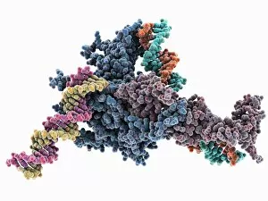
Chromatin remodelling factor and DNA, molecular model. The strands of DNA (deoxyribonucleic acid) are at left and right (pink-yellow and green-orange). This chromatin remodelling factor is ISW1a

Background imageOncology Collection: Tumour embolism, light micrograph C015 / 6397

Tumour embolism, light micrograph C015 / 6397
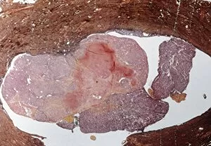
Tumour embolism. Light micrograph of a section through an embolism (blood clot) caused by cells from a cervical cancer. The cells have left the primary tumour and are metastasising (spreading)

Background imageOncology Collection: Mesothelioma lung cancer, X-ray C017 / 7776

Mesothelioma lung cancer, X-ray C017 / 7776
Mesothelioma lung cancer, X-ray of the chest of a 84 year old male patient with advanced mesothelioma, a type of lung cancer

Background imageOncology Collection: Angiosarcoma of the spleen C015 / 6398

Angiosarcoma of the spleen C015 / 6398
Angiosarcoma. Gross specimen of a sectioned spleen affected by angiosarcoma. Malignant (cancerous) tumours (yellow) can be seen at bottom centre


All Professionally Made to Order for Quick Shipping

Oncology: Unveiling the Battle Within In the realm of oncology, a fierce war is waged between T lymphocytes and cancer cells. SEM C001 / 1679 captures this epic struggle at a microscopic level, showcasing the intricate dance between these two adversaries. HeLa cells, depicted in light micrographs C017 / 8299 and C017 / 8298, offer us glimpses into their mysterious world. These immortal cells have played an instrumental role in countless scientific breakthroughs, unraveling the secrets of cancer biology. Acute promyelocytic leukemia reveals its sinister face through a haunting micrograph. The image serves as a reminder of the relentless fight against blood cancers that oncologists tirelessly engage in each day. Ovarian cancer's treacherous nature is exposed under the lens of light micrograph C015 / 7103. This glimpse into its cellular landscape fuels our determination to develop effective treatments for this formidable disease. The paclitaxel drug molecule emerges as a potent weapon in our arsenal against cancer. Its molecular structure holds promise for patients battling various forms of malignancies. Within operating rooms worldwide, surgeons skillfully navigate breast cancer cases with precision and expertise. Armamentarii Chirurgici provides us with an intimate view into these life-saving procedures. Cancerous scrofula reminds us that skin diseases can also be harbingers of deeper battles within our bodies. Description des Maladies de la Peau offers insight into dermatological manifestations linked to underlying malignancies. Traite Complet de l'Anatomie de l unveils surgical interventions on malignant tumors – visual proof that medical professionals are at the forefront of combating this pervasive disease. Medical illustrations depicting pilonidal cysts near the natal cleft or stages of colon cancer serve as educational tools to raise awareness about lesser-known aspects while empowering individuals to seek timely medical intervention.