Fraud Blocker Skip to main content

Organelle Collection (page 6)

Organelles: The Tiny Powerhouses Within Our Cells The world of cells is a fascinating one, filled with intricate structures known as organelles

Background imageOrganelle Collection: Animal cell organelles, artwork C016 / 0610

Animal cell organelles, artwork C016 / 0610
Computer artwork showing the organelles in a eukaryotic cell. This is an animal cell. Structures include the nucleus (centre, orange) which has a membrane with nuclear pores
Background imageOrganelle Collection: Animal cell organelles, artwork C016 / 0604

Animal cell organelles, artwork C016 / 0604
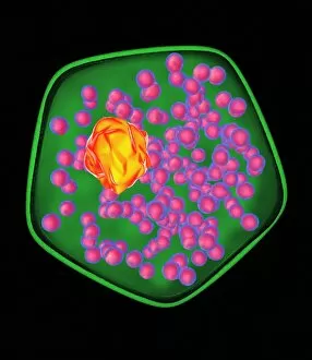
Computer artwork showing the organelles in a eukaryotic cell. This is an animal cell. Structures include the nucleus (centre, red) which has a membrane with nuclear pores

Background imageOrganelle Collection: Animal cell organelles, artwork C016 / 0606

Animal cell organelles, artwork C016 / 0606
Computer artwork showing the organelles in a eukaryotic cell. This is an animal cell. Structures include the nucleus (centre, orange) which has a membrane with nuclear pores

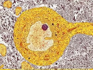
Background imageOrganelle Collection: Purkinje nerve cell, TEM C014 / 0582

Purkinje nerve cell, TEM C014 / 0582
Purkinje nerve cell. Transmission electron micrograph (TEM) of a purkinje nerve cell (bright yellow, centre) from the cerebellum of the brain

Background imageOrganelle Collection: Chloroplast, TEM C017 / 8233

Chloroplast, TEM C017 / 8233
Chloroplast. Coloured transmission electron micrograph (TEM) of chloroplast from the leaf of a Coleus blumei plant. Chloroplasts are the sites of photosynthesis

Background imageOrganelle Collection: Islet of Langerhans, TEM C015 / 6418

Islet of Langerhans, TEM C015 / 6418
Islet of Langerhans. Transmission electron micrograph (TEM) of a section through cells clustered in an islet of Langerhans in the pancreas

Background imageOrganelle Collection: Fibroblast cell, fluorescent micrograph

Fibroblast cell, fluorescent micrograph
Fibroblast cell. Confocal laser scanning micrograph of a fibroblast cell. The cell nucleus, which contains the cells genetic information, is green

Background imageOrganelle Collection: Plant protoplast, fluorescent micrograph

Plant protoplast, fluorescent micrograph
Protoplast. Confocal laser scanning micrograph of a tobacco plant (Nicotiana tabacum) protoplast. A protoplast is a plant cell that has had its tough outer cell wall removed by chemical treatment

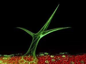
Background imageOrganelle Collection: Plant trichome, fluorescent micrograph

Plant trichome, fluorescent micrograph
Plant trichome. Confocal laser scanning micrograph of a trichome (leaf hair) on a Arabidopsis thaliana plant. Actin filaments in the trichome are green

Background imageOrganelle Collection: Animal cell, artwork C013 / 9985

Animal cell, artwork C013 / 9985
Animal cell. Computer artwork showing the cell organelles found inside a typical animal cell. The nucleus (large round) can be seen at centre

Background imageOrganelle Collection: Animal cell, artwork C013 / 9984

Animal cell, artwork C013 / 9984
Animal cell. Computer artwork showing the cell organelles found inside a typical animal cell. The nucleus (large round) can be seen at centre

Background imageOrganelle Collection: Human cell, artwork C013 / 6522

Human cell, artwork C013 / 6522
Human cell. Illustration of a section through a human cell. At centre is the nucleus (purple), with the nucleolus (dark red) inside it

Background imageOrganelle Collection: Sperm cell anatomy, artwork C013 / 4648

Sperm cell anatomy, artwork C013 / 4648
Sperm cell anatomy. Cutaway computer artwork showing the internal structure of a sperm cell (spermatozoon), the male sex cell

Background imageOrganelle Collection: Intracellular transport, artwork C013 / 4997

Intracellular transport, artwork C013 / 4997
Intracellular transport. Computer artwork of vesicles (spheres) being transported from a Golgi body (blue, left) around the cell by microtubules (string-like)

Background imageOrganelle Collection: Mitochondria, artwork C013 / 4993

Mitochondria, artwork C013 / 4993
Mitochondria, computer artwork. Mitochondria are a type of organelle found in the cytoplasm of eukaryotic cells. They oxidise sugars and fats to produce energy in a process called respiration

Background imageOrganelle Collection: Intracellular transport, artwork C013 / 4995

Intracellular transport, artwork C013 / 4995
Intracellular transport. Computer artwork of vesicles (spheres) being transported from a Golgi body (blue, top) around the cell by microtubules (string-like)

Background imageOrganelle Collection: Mitochondria, artwork C013 / 4991

Mitochondria, artwork C013 / 4991
Mitochondria, computer artwork. Mitochondria are a type of organelle found in the cytoplasm of eukaryotic cells. They oxidise sugars and fats to produce energy in a process called respiration

Background imageOrganelle Collection: Nerve cell, TEM C013 / 4797

Nerve cell, TEM C013 / 4797
Nerve cell. Transmission electron micrograph (TEM) of a section through a neuron (nerve cell), showing characteristic Nissl body (dark blue lines), numerous golgi apparatus (curved green lines)

Background imageOrganelle Collection: Nerve cell, TEM C013 / 4796

Nerve cell, TEM C013 / 4796
Nerve cell. Transmission electron micrograph (TEM) of a section through a neuron (nerve cell), showing characteristic Nissl body (dark red lines), numerous golgi apparatus (curved pink lines)

Background imageOrganelle Collection: Pancreatic cells, TEM

Pancreatic cells, TEM
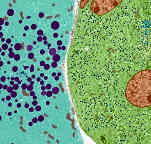
Pancreatic cells. Coloured transmission electron micrograph (TEM) of an acinar (exocrine) pancreatic cell (blue) adjacent to an hormone- secreting (endocrine) Islet of Langerhans cell (green)

Background imageOrganelle Collection: Pancreatic cell, TEM

Pancreatic cell, TEM
Pancreatic cell. Coloured transmission electron micrograph (TEM) of part of an acinar (exocrine) pancreatic cell. Mitochondria (red) are seen in the cells endoplasmic reticulum (yellow)

Background imageOrganelle Collection: Pancreatic alpha cell, TEM

Pancreatic alpha cell, TEM
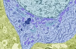
Pancreatic alpha cell. Coloured transmission electron micrograph (TEM) of a hormone-secreting (endocrine) alpha cell, found in the islets of Langerhans of the pancreas

Background imageOrganelle Collection: Cardiac muscle

Cardiac muscle. Coloured scanning electron micrograph (SEM) of heart (cardiac) muscle fibrils (yellow). The membrane around the muscle has been torn (yellow)

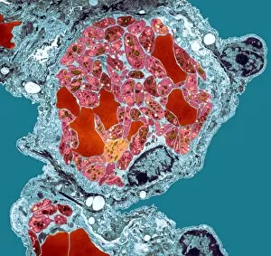
Background imageOrganelle Collection: Blood platelet, TEM

Blood platelet, TEM

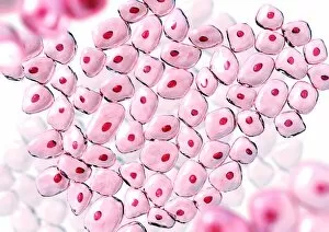
Background imageOrganelle Collection: Cells

Cells. Computer artwork of non-specific cells. The red dot in the centre of each cell is the nucleus where the DNA (deoxyribonucleic acid) of each cell is stored

Background imageOrganelle Collection: Golgi apparatus, TEM

Golgi apparatus, TEM
Golgi apparatus. Coloured transmission electron micrograph (TEM) of Golgi apparatus, also known as Golgi bodies, (pink) in a macrophage white blood cell

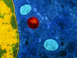
Background imageOrganelle Collection: Colour TEM of primary lysosome in liver cell

Colour TEM of primary lysosome in liver cell
Primary lysosome. Coloured Transmission Electron Micrograph (TEM) of a primary lysosome seen in a liver cell. The lysosome (red) is a membrane bound structure that contains digestive enzymes

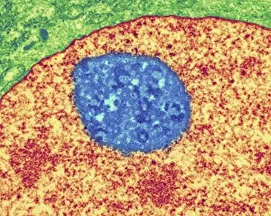
Background imageOrganelle Collection: Cell nucleolus, TEM

Cell nucleolus, TEM
Nucleolus. Coloured transmission electron micrograph of the nucleolus (blue), a component of the nucleus (orange) of a cell. The cell cytoplasm is green

Background imageOrganelle Collection: Animal cell structure, computer artwork

Animal cell structure, computer artwork
Animal cell. Computer artwork of a section through an animal cell. At the centre is the nucleus, which contains the cells genetic information in the form of DNA (deoxyribonucleic acid)

Background imageOrganelle Collection: Cell organelles, TEM

Cell organelles, TEM
Cell organelles. Coloured transmission electron micrograph (TEM) of several organelles in an animal cell. At centre, the nucleus (dark purple, round) contains the cells genetic material (DNA)

Background imageOrganelle Collection: Mitochondria

Mitochondria. Coloured transmission electron micrograph of mitochondria (circles). A mitochondrion is a membrane-bound organelle that exists in eukaryotic cells

Background imageOrganelle Collection: Cellular clock

Cellular clock. Conceptual artwork of a clock and the internal structures (organelles) of an animal cell. This could represent the rate of metabolic cell reactions

Background imageOrganelle Collection: Evolving protocell, artwork

Evolving protocell, artwork
Evolving protocell. Image 3 of 5. Artwork showing a protocell (artificial cell). This protocell was formed from a synthetic primeval soup containing PNA (peptide nucleic acid, orange)

Background imageOrganelle Collection: Ribosomes, TEM

Ribosomes, TEM
Ribosomes. Coloured transmission electron micrograph (TEM) of ribosomes (blue) passing through pores in a cells nuclear membrane (red)

Background imageOrganelle Collection: Protocell proliferation, artwork

Protocell proliferation, artwork
Protocell proliferation. Image 5 of 5. Artwork showing a protocell (artificial cell) dividing to produce two daughter cells

Background imageOrganelle Collection: Cardiac muscle, SEM

Cardiac muscle, SEM
Cardiac muscle. Coloured scanning electron micrograph (SEM) of a bundle of cardiac muscle fibrils (green) from a healthy heart. Mitochondria (round, orange) supply the muscle cells with energy

Background imageOrganelle Collection: Mitochondrial Eve

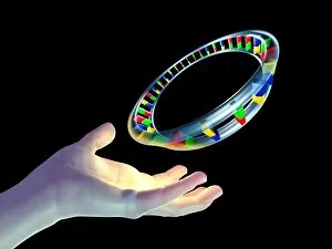
Mitochondrial Eve. Conceptual computer artwork of a circular molecule of mitochondrial deoxyribonucleic acid (DNA) floating above a hand. Mitochondria are cell organelles that produce energy

Background imageOrganelle Collection: Newly formed protocell, artwork

Newly formed protocell, artwork
Newly formed protocell. Image 2 of 5. Artwork showing cellular components inside a basic cell membrane. This protocell (artificial cell)

Background imageOrganelle Collection: Energised protocell, artwork

Energised protocell, artwork
Energised protocell. Image 4 of 5. Artwork showing light photons energising a protocell (artificial cell). This protocell was formed from a synthetic primeval soup containing PNA

Background imageOrganelle Collection: Plant cells, artwork

Plant cells, artwork
Plant cells, computer artwork

Background imageOrganelle Collection: Plant cell, artwork

Plant cell, artwork
Plant cell, computer artwork


All Professionally Made to Order for Quick Shipping

Organelles: The Tiny Powerhouses Within Our Cells The world of cells is a fascinating one, filled with intricate structures known as organelles. These tiny entities play crucial roles in maintaining the functionality and survival of our cells. From budding yeast to HeLa cells, each cell type possesses its unique set of organelles that contribute to their specialized functions. Under the lens of a light microscope, the HeLa cells reveal mesmerizing patterns resembling abstract artwork. Their delicate structures are visible, showcasing the complexity within these microscopic powerhouses. Meanwhile, another image captures nerve cells in all their glory - long and slender extensions reaching out like branches from a tree. Zooming in further using transmission electron microscopy (TEM), we get an up-close look at some specific organelles. The rough endoplasmic reticulum appears as a network of interconnected membranes studded with ribosomes responsible for protein synthesis. Mitochondria steal the spotlight next; these bean-shaped powerhouses generate energy for cellular activities through respiration. But it's not just animal cells that possess remarkable organelles; plant cells have their own unique features too. Chloroplasts, depicted beautifully through artwork, capture sunlight and convert it into energy via photosynthesis—a process vital for sustaining life on Earth. Another TEM image reveals Purkinje nerve cells—large neurons found in our brain's cerebellum—showcasing their intricate branching structure responsible for coordinating movement and balance. Finally, scanning electron microscopy (SEM) unveils the Golgi apparatus—an organelle involved in processing proteins and packaging them into vesicles for transportation throughout the cell or secretion outside it. These glimpses into different types of organelles highlight their diverse forms and functions within our cellular world. They remind us that even though they may be invisible to the naked eye, these minuscule entities hold immense importance in keeping our bodies functioning harmoniously at every level.