
Osteoblast Collection
Osteoblasts, the architects of healthy bone, work tirelessly to build and maintain our skeletal system


All Professionally Made to Order for Quick Shipping
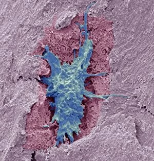
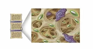
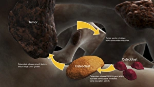
Osteoblasts, the architects of healthy bone, work tirelessly to build and maintain our skeletal system. These remarkable cells are responsible for synthesizing and depositing new bone tissue, ensuring its strength and integrity. Under the scanning electron microscope (SEM), we can observe the intricate structure of osteocytes - mature bone cells that reside within their own lacunae. Their long dendritic processes extend through tiny channels called canaliculi, forming a network that allows them to communicate with neighboring cells. In contrast, osteoclasts play a different role in maintaining bone health. These specialized cells possess an impressive ability to break down old or damaged bone tissue through a process known as resorption. SEM images capture these powerful osteoclasts at work, eroding bone in conditions like osteoporosis or during the natural remodeling process. Bone metastasis is another phenomenon captured conceptually; it illustrates how cancerous cells spread from other parts of the body into bones, disrupting their normal function. This image serves as a reminder of the importance of early detection and treatment for such conditions. Light micrographs provide us with detailed views of various aspects related to bones: from growth patterns seen in developing skeletons to structural units that make up our skeleton's framework. Lastly, transmission electron microscopy (TEM) offers an even closer look at osteoblasts' ultrastructure - revealing their organelles involved in protein synthesis and mineralization processes. These TEM images highlight just how intricately designed these cells are for building strong bones.