Fraud Blocker Skip to main content

Osteology Collection

Osteology, the fascinating study of bones and skeletal structures, unveils a world of intricate details and medical advancements

Background imageOsteology Collection: Skeletons, X-ray artwork

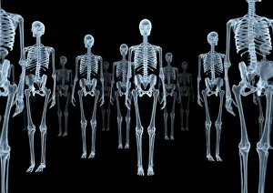
Skeletons, X-ray artwork
Skeleton crowd. Computer X-ray artwork of a group of human skeletons viewed from the front

Background imageOsteology Collection: Running skeleton in body, artwork

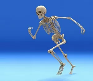
Running skeleton in body, artwork
Running skeleton inside an outline of the body, computer artwork

Background imageOsteology Collection: Headache, X-ray artwork

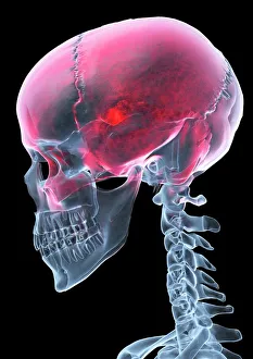
Headache, X-ray artwork
Headache. Computer X-ray artwork of a lateral view of a human skull. The red area represents pain caused by a headache (cephalalgia)

Background imageOsteology Collection: Total hip replacement, X-ray

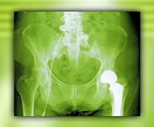
Total hip replacement, X-ray
Total hip replacement. Coloured frontal X-ray of a female pelvis with a total hip replacement (white, lower right)

Background imageOsteology Collection: Fractured ankle, X-ray

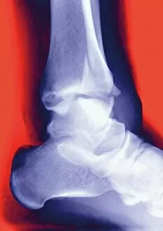
Fractured ankle, X-ray
Fractured ankle. Coloured profile X-ray of a distal fibula fracture (upper centre). The fibula is the smaller leg bone running down from top centre. The larger bone is the tibia

Background imageOsteology Collection: Calots spinal surgery, 19th century

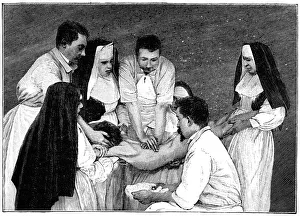
Calots spinal surgery, 19th century
Calots spinal surgery, 19th-century artwork. This operation is being carried out by the French surgeon Jean-Francois Calot (1861-1944) on a condition known as Potts disease

Background imageOsteology Collection: Damaged knee ligament, artwork

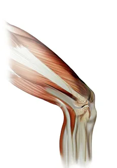
Damaged knee ligament, artwork
Damaged knee ligament. Computer artwork of the muscle structure of the leg and anatomy of the human knee joint, showing a tear (dark line, centre right) in one of the cruciate ligaments (beige)

Background imageOsteology Collection: Skeleton from below, X-ray artwork

Skeleton from below, X-ray artwork
Skeleton. Computer x-ray artwork of a human skeleton viewed from below

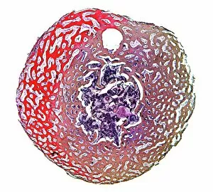
Background imageOsteology Collection: Compact bone, light micrograph

Compact bone, light micrograph
Compact bone. Polarised light micrograph of a transverse section through compact bone tissue, showing Haversian canals (circular regions)

Background imageOsteology Collection: Osteocyte bone cell, SEM C016 / 9025

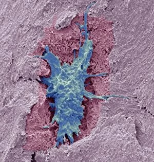
Osteocyte bone cell, SEM C016 / 9025
Osteocyte bone cell. Coloured scanning electron micrograph (SEM) of an osteocyte bone cell (blue) surrounded by bone tissue (pink)

Background imageOsteology Collection: Normal spine, X-ray

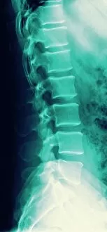
Normal spine, X-ray
Normal spine. Coloured profile X-ray showing bones of the spine (vertebrae, centre). The front of the body is at right. The ribs (upper right) and the pelvis (bottom) are also seen

Background imageOsteology Collection: Spine with workers, spine repair F007 / 9907

Spine with workers, spine repair F007 / 9907
Conceptual computer artwork depicting a spine being repaired

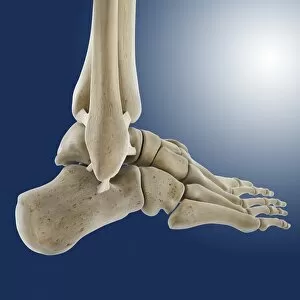
Background imageOsteology Collection: Outer ankle ligaments, artwork C013 / 4452

Outer ankle ligaments, artwork C013 / 4452
Outer ankle ligaments. Computer artwork of the bones and ligaments (white) of the right foot and ankle seen from the side

Background imageOsteology Collection: Fractured jawbone, X-ray

Fractured jawbone, X-ray
Fractured jawbone. Coloured X-ray of a human jaw showing a fractured lower jawbone (mandible). The fracture has occurred to the left side of the lower jawbone (bottom right, orange)

Background imageOsteology Collection: Inner ankle ligaments, artwork C013 / 4451

Inner ankle ligaments, artwork C013 / 4451
Inner ankle ligaments. Computer artwork of the bones and ligaments (white) of the right foot and ankle seen from the side

Background imageOsteology Collection: Spongy bone, light micrograph

Spongy bone, light micrograph
Spongy bone. Light micrograph of a section through stained and decalcified human spongy bone. This bone type is also called cancellous bone

Background imageOsteology Collection: Human Back and Buttock Muscles, Posterior, 1844

Human Back and Buttock Muscles, Posterior, 1844

Background imageOsteology Collection: Bones of the skeleton, from Tabulae Osteologicae

Bones of the skeleton, from Tabulae Osteologicae
CHT235091 Bones of the skeleton, from Tabulae Osteologicae by Christoph Jacob Trew (1695-1769) engraved by G

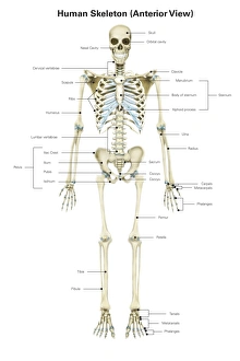
Background imageOsteology Collection: Anterior view of human skeletal system, with labels

Anterior view of human skeletal system, with labels

Background imageOsteology Collection: Anatomy of human skull from different angles

Anatomy of human skull from different angles

Background imageOsteology Collection: Human skeletal system, front view

Human skeletal system, front view

Background imageOsteology Collection: Normal legs, X-rays

Normal legs, X-rays
Normal legs. Coloured frontal X-rays of the healthy legs, of a 35-year-old

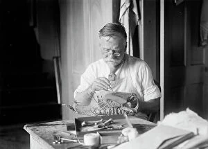
Background imageOsteology Collection: J. W. Scollick of Smithsonian Institute

J. W. Scollick of Smithsonian Institute, 1916. Special osteological preparator at U.S. National Museum

Background imageOsteology Collection: Bones of the foot, from Tabulae Osteologicae by Christoph Jacob Trew (1695-1769

Bones of the foot, from Tabulae Osteologicae by Christoph Jacob Trew (1695-1769
CHT235090 Bones of the foot, from Tabulae Osteologicae by Christoph Jacob Trew (1695-1769) engraved by G

Background imageOsteology Collection: Side view of human skull with parts exploded

Side view of human skull with parts exploded

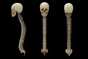
Background imageOsteology Collection: 3D rendering of human vertebral column with skull

3D rendering of human vertebral column with skull

Background imageOsteology Collection: Male human head with skull in ghost effect, side view

Male human head with skull in ghost effect, side view. on white background

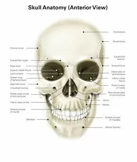
Background imageOsteology Collection: Anterior view of human skull, with labels

Anterior view of human skull, with labels

Background imageOsteology Collection: Anatomy of human pelvic bone

Anatomy of human pelvic bone

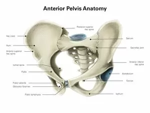
Background imageOsteology Collection: Anterior view of human pelvis, with labels

Anterior view of human pelvis, with labels

Background imageOsteology Collection: Male human skeleton in dynamic posture, front view

Male human skeleton in dynamic posture, front view

Background imageOsteology Collection: Anatomy of human skull, side view

Anatomy of human skull, side view

Background imageOsteology Collection: Posterior view of left hand, with labels

Posterior view of left hand, with labels

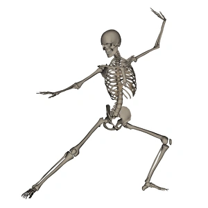
Background imageOsteology Collection: Front view of human skeleton in fighting stance

Front view of human skeleton in fighting stance, isolated on white background

Background imageOsteology Collection: Front view of a human skeleton posing

Front view of a human skeleton posing, isolated on white background

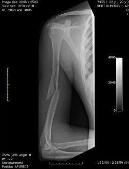
Background imageOsteology Collection: Broken arm bone, digital X-ray

Broken arm bone, digital X-ray
Broken arm bone. Digital frontal X-ray of the fractured right humerus (upper arm bone) of a patient in their early twenties

Background imageOsteology Collection: Normal hand, digital X-ray

Normal hand, digital X-ray
Normal hand. Digital X-ray of the normal right hand of a patient in their late twenties. Various sets of data relating to the image can be seen around the edges of the X-ray

Background imageOsteology Collection: Human skeleton anatomy, artwork

Human skeleton anatomy, artwork
Human skeleton anatomy, computer artwork. Three views of the bones of the human torso, seen from an oblique frontal view (left), from the side (centre) and from the front (right)

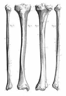
Background imageOsteology Collection: Leg bones anatomy engraving 1866

Leg bones anatomy engraving 1866
Atlas d anatomie descriptive du corps humain C. Bonamy - Paul Broca Victor Masson et Fils Paris 1866

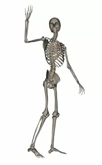
Background imageOsteology Collection: Front view of human skeleton waving goodbye

Front view of human skeleton waving goodbye, isolated on white background

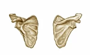
Background imageOsteology Collection: Medical illustration of human scapula bone

Medical illustration of human scapula bone

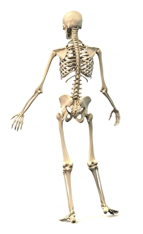
Background imageOsteology Collection: Male human skeleton in dynamic posture, rear view

Male human skeleton in dynamic posture, rear view

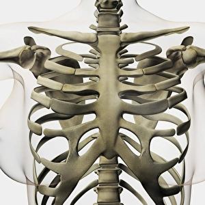
Background imageOsteology Collection: Three dimensional view of female sternum and rib cage

Three dimensional view of female sternum and rib cage

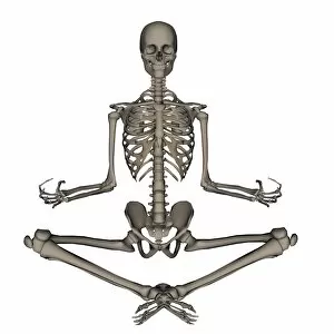
Background imageOsteology Collection: Front view of human skeleton meditation

Front view of human skeleton meditation, isolated on white background

Background imageOsteology Collection: Anterior view of human skeletal system

Anterior view of human skeletal system

Background imageOsteology Collection: Perspective view of human skull

Perspective view of human skull with parts exploded

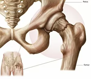
Background imageOsteology Collection: Anatomy of hip fracture

Anatomy of hip fracture

Background imageOsteology Collection: X-ray image of knee

X-ray image of knee

Background imageOsteology Collection: Three dimensional view of female skeletal system

Three dimensional view of female skeletal system

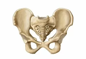
Background imageOsteology Collection: Three dimensional view of human pelvic bones

Three dimensional view of human pelvic bones

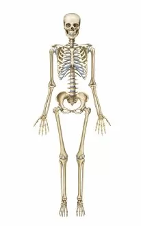
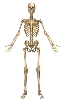
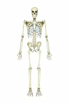
Background imageOsteology Collection: Front view of human skeleton

Front view of human skeleton, isolated on white background

Background imageOsteology Collection: Front view of human skeleton running

Front view of human skeleton running, isolated on white background

Background imageOsteology Collection: Front view of human skeleton meditating

Front view of human skeleton meditating, isolated on white background

Background imageOsteology Collection: Temporomandibular joint, normal and dislocated

Temporomandibular joint, normal and dislocated

Background imageOsteology Collection: The human skeleton and muscular system, front view

The human skeleton and muscular system, front view
Medical illustration of the human skeleton and muscular system, front view

Background imageOsteology Collection: Human skeleton praying on his knees

Human skeleton praying on his knees, isolated on white background

Background imageOsteology Collection: Three dimensional view of female rib cage and skeletal system

Three dimensional view of female rib cage and skeletal system

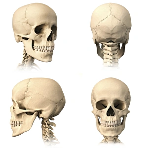
Background imageOsteology Collection: View of human skull from different angles

View of human skull from different angles

Background imageOsteology Collection: Hip joint bones and anatomy, artwork C014 / 2032

Hip joint bones and anatomy, artwork C014 / 2032
Hip joint bones and anatomy. Artwork of the bones that form the two hip joints (right and left). The hip is a ball-and-socket joint that provides the mobility needed for walking and running

Background imageOsteology Collection: Normal knees, X-rays

Normal knees, X-rays
Normal knees. Coloured frontal X-rays of the healthy knees of a 35-year-old

Background imageOsteology Collection: Thickened skull, MRI scan

Thickened skull, MRI scan
Thickened skull. Magnetic resonance imaging (MRI) scan of an axial section through the head of a 22-year-old patient, showing thickening of the trabecular (spongy) bone of the skull

Background imageOsteology Collection: Total knee replacement, X-ray

Total knee replacement, X-ray
Total knee replacement. X-ray of the legs of a 49-year-old patient who has had a total knee replacement (white) of the left knee (right)

Background imageOsteology Collection: Osteocyte bone cell, SEM C016 / 9026

Osteocyte bone cell, SEM C016 / 9026
Osteocyte bone cell. Coloured scanning electron micrograph (SEM) of an osteocyte bone cell (red) surrounded by bone tissue (grey)

Background imageOsteology Collection: Paprosky femur defect classification C016 / 6621

Paprosky femur defect classification C016 / 6621
Paprosky femur defect classification. Diagram showing the classification system for femur cortex defects used to assess revision (replacement or repair) of a hip implant

Background imageOsteology Collection: Bone tissue

Bone tissue. Coloured scanning electron micrograph (SEM) of cancellous (spongy) bone. Bone tissue can be either cortical (compact) or cancellous

Background imageOsteology Collection: Kneecap fracture, X-ray

Kneecap fracture, X-ray
Kneecap fracture. Coloured X-ray of a broken kneecap (patella), as viewed from the side with the knee bent. The kneecap (far left, centre) is a disc of bone that covers the front of the knee joint

Background imageOsteology Collection: Fractured foot, coloured X-ray

Fractured foot, coloured X-ray
Fractured foot. Coloured X-rays of a fractured fifth metatarsal (to the right of each image). The fracture is seen as a small black crack

Background imageOsteology Collection: Healthy skull, coloured X-ray

Healthy skull, coloured X-ray
Healthy human skull. Side view X-ray showing the skull and the outline of the head (black). The bones of the cranium are fused together to form a complete structure that protects the brain

Background imageOsteology Collection: Normal lumbar spine, X-ray

Normal lumbar spine, X-ray
Normal lumbar spine. Coloured frontal X-ray showing bones of the lower (lumbar) spine (centre, purple). The bones of the spine (block-shaped) are called vertebrae

Background imageOsteology Collection: Neck and shoulder pain, X-ray artwork

Neck and shoulder pain, X-ray artwork
Neck and shoulder pain. Computer X-ray artwork of the back view of a human skeletons shoulders, neck and skull. The red area represents neck pain (cervicalgia) and shoulder pain

Background imageOsteology Collection: Normal female pelvis, X-ray

Normal female pelvis, X-ray
Normal female pelvis, coloured frontal X-ray. The thigh (femur) bones of the leg are seen at bottom left and right, and the base of the spine is at top

Background imageOsteology Collection: Osteoblast bone cell, SEM

Osteoblast bone cell, SEM
Osteoblast bone cell. Coloured scanning electron micrograph (SEM) of an osteoblast (centre) in bone tissue. Osteoblasts are primarily found in regions of new bone growth

Background imageOsteology Collection: Whale bone tissue, light micrograph

Whale bone tissue, light micrograph

Background imageOsteology Collection: Skeleton lying down, artwork

Skeleton lying down, artwork
Skeleton lying down. Computer artwork showing the bones of the head, neck and upper torso of a human skeleton that is lying down

Background imageOsteology Collection: Anterior view of human skull

Anterior view of human skull

Background imageOsteology Collection: Conceptual image of human skull, side view

Conceptual image of human skull, side view

Background imageOsteology Collection: Human skeletal system

Human skeletal system

Background imageOsteology Collection: Medical illustration of a womans skull and skeletal system

Medical illustration of a womans skull and skeletal system, three dimensional view

Background imageOsteology Collection: Front view of human skeletal system

Front view of human skeletal system

Background imageOsteology Collection: Anatomy of human vertebra

Anatomy of human vertebra

Background imageOsteology Collection: 3D rendering of human teeth and skull

3D rendering of human teeth and skull

Background imageOsteology Collection: 3D rendering of human skeletal system, rear view

3D rendering of human skeletal system, rear view

Background imageOsteology Collection: Illustration of knee bone, bending

Illustration of knee bone, bending

Background imageOsteology Collection: Conceptual image of pain in human foot

Conceptual image of pain in human foot


All products are expertly crafted, using premium materials, tailored to your specifications and promptly shipped

Osteology, the fascinating study of bones and skeletal structures, unveils a world of intricate details and medical advancements. From total hip replacements to X-ray imaging, this field has revolutionized the way we understand and treat bone-related conditions. In the realm of orthopedics, total hip replacement surgeries have become a game-changer for those suffering from debilitating joint pain. With cutting-edge techniques and prosthetic materials, these procedures restore mobility and improve quality of life. Delving into history, Calots spinal surgery in the 19th century stands as a testament to human resilience. Despite limited resources and knowledge at that time, surgeons courageously ventured into complex spinal operations with remarkable outcomes. Artwork captures both the beauty and vulnerability of our skeletal system. A damaged knee ligament depicted in brushstrokes reminds us of how delicate our bodies can be. Meanwhile, a running skeleton portrayed through art showcases strength and agility even in its bare form. X-ray artwork takes us beneath the surface to reveal hidden stories within our bones. Skeletons captured by this technique offer an ethereal glimpse into their structure while evoking curiosity about their past lives. An X-ray artwork showcasing a skeleton from below adds another dimension to our understanding of anatomy. It invites contemplation on how every angle tells a different story about our body's framework. Sometimes even mundane ailments find artistic expression through X-rays. A headache immortalized as an artwork serves as a reminder that even seemingly minor discomforts can leave traces within us. Fractured ankle X-rays remind us that bones are not invincible; they can break under pressure or unfortunate circumstances. Yet with modern medicine's intervention, healing is possible once again. Microscopic exploration reveals wonders too small for the naked eye to behold. Compact bone viewed under light micrograph exposes its intricate structure like never before – reminding us that there is more than meets the eye when it comes to osteology.

© Copyright Media Storehouse, All Rights Reserved 2003 - 2024
Facebook     Pinterest     100% Payment Secure     Reviews IO