
Otology Collection
Otology, the fascinating field of study dedicated to the intricate anatomy and physiology of the human ear, has a rich history that dates back centuries


All Professionally Made to Order for Quick Shipping
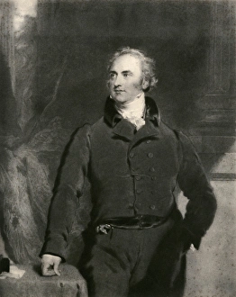
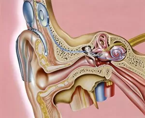
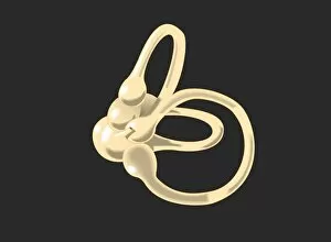
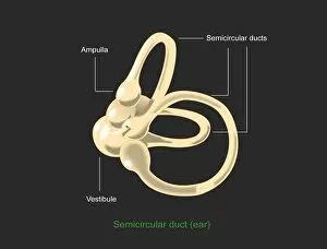
Otology, the fascinating field of study dedicated to the intricate anatomy and physiology of the human ear, has a rich history that dates back centuries. Pioneers like Sir Astley Paston Cooper paved the way for our understanding of this complex sensory organ. In the 19th century, detailed engravings emerged as powerful tools in otological research. An anatomical representation of the human ear canal captured by American engravers showcased its delicate structure and provided valuable insights into its functioning. Similarly, French engravers contributed with their own depictions of both the ear canal and middle ear, unraveling further mysteries surrounding this remarkable system. These engravings were accompanied by a series of diagrams illustrating different parts of the human ear. Each diagram meticulously highlighted crucial components such as cochlea implantation sites and Eustachian tube anatomy. These visual aids allowed researchers to delve deeper into understanding hearing mechanisms and develop innovative solutions for auditory impairments. One such groundbreaking invention was the cochlear implant depicted in artwork C016/7683. This technological marvel revolutionized otology by providing hope to individuals with severe hearing loss or deafness. By bypassing damaged hair cells within the inner ear, these implants directly stimulate auditory nerves, enabling recipients to perceive sound once again. Moreover, studies on primate ears conducted through scanning electron microscopy (SEM) shed light on similarities between humans and our closest relatives in terms of their intricate ear canals. These findings not only deepen our knowledge about evolution but also aid in developing treatments for various auditory disorders affecting primates. Furthermore, SEM images showcasing semicircular canals emphasize how balance sensing is intricately connected to otology. Understanding these structures helps us comprehend equilibrium-related conditions like vertigo more comprehensively while paving new avenues for treatment options. As we continue exploring otology's vast realm through artistry and scientific advancements alike, we unlock countless possibilities for improving hearing health worldwide.