
Palps Collection
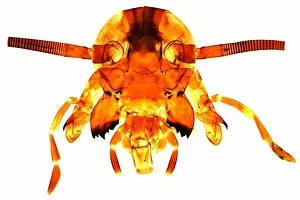
"Exploring the Intricate World of Palps: A Closer Look at Tick Mouthparts and Fly Heads" In this captivating journey through the microscopic world


All Professionally Made to Order for Quick Shipping
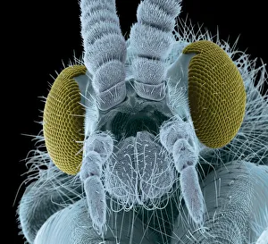
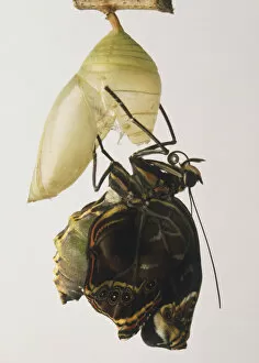
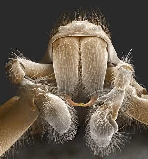
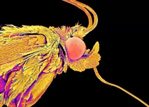
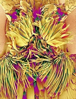
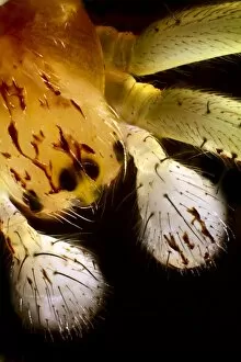
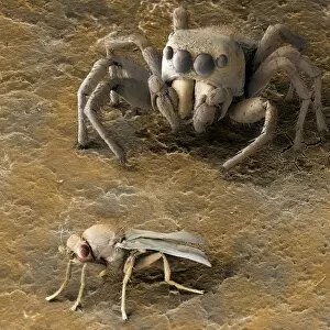
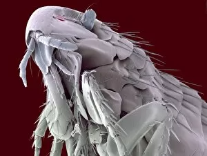
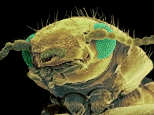
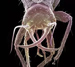
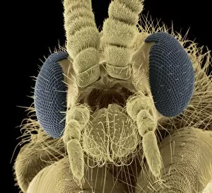
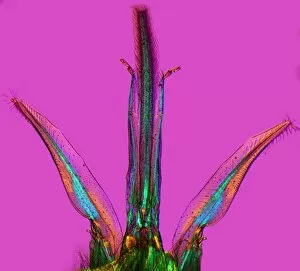
"Exploring the Intricate World of Palps: A Closer Look at Tick Mouthparts and Fly Heads" In this captivating journey through the microscopic world, we delve into the fascinating realm of palps. These tiny structures, found in various creatures such as ticks and flies, play a crucial role in their survival. Through scanning electron microscopy (SEM), we are granted an up-close view of a fly's head. The colored scanning electron micrograph reveals intricate details, highlighting the complexity of these remarkable organisms. Meanwhile, high up in Kleine Scheidegg, Switzerland, Hotel Bellevue - Jungfrau stands majestically against a breathtaking backdrop – reminding us that nature's wonders can be found even amidst human creations. Zooming in further on our exploration, we encounter the Giant House Spider (Tegenaria gigantea) adult male. Its close-up reveals not only its fearsome appearance but also showcases its impressive head and palps. Similarly enchanting is witnessing the emergence of a Blue Morpho butterfly from its cocoon – an awe-inspiring transformation captured with vibrant colors. However, not all creatures evoke wonder; some trigger fear instead. The Black Australian funnelweb spider spinning its trap sends shivers down our spines when viewed up close. Yet even amongst arachnids lies beauty – like a Jumping spider perched upon a rock with intense focus or observing a hairy brown spider with four eyes staring back at us curiously. As our journey continues to unfold before our eyes, we come across more spiders – each unique in their appearance and behavior. From hairy black spiders lurking on the ground to striking black and yellow ones with arched legs poised for action; they remind us that diversity exists even within seemingly similar species. Finally, SEM allows us to marvel at spider mouthparts under Z430 / 0436 magnification – revealing intricacies hidden from plain sight yet vital for their survival.