
Parenchyma Collection (page 2)
Parenchyma is a vital tissue found in various parts of plants, playing essential roles in their growth and development


All Professionally Made to Order for Quick Shipping
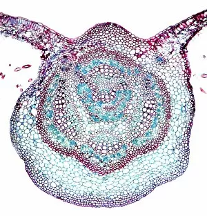
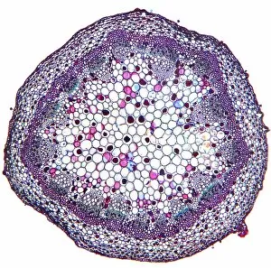
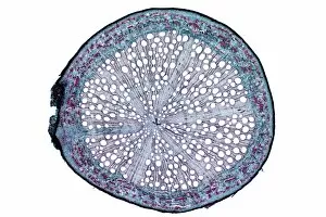
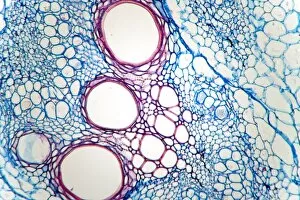
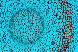
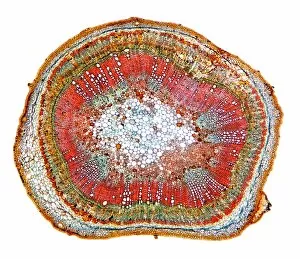
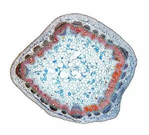
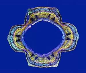
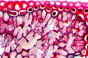
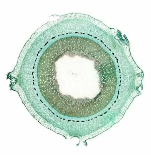
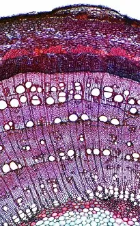
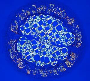
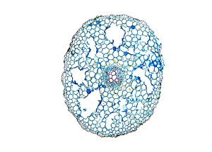
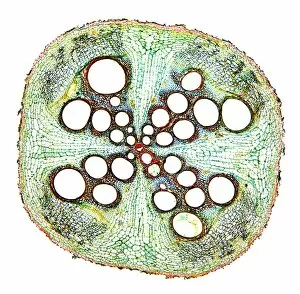
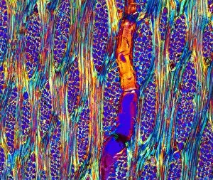
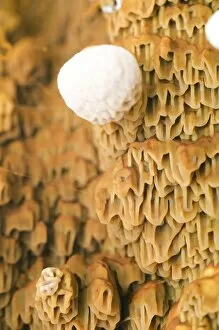
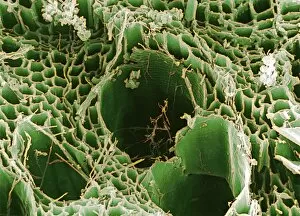
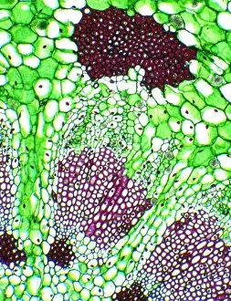
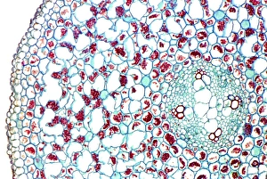
Parenchyma is a vital tissue found in various parts of plants, playing essential roles in their growth and development. From the pine stem to the castor oil stem, parenchyma cells can be observed under a light microscope, revealing their unique structures and functions. In the light micrograph of a pine stem, clusters cells can be seen scattered throughout the tissue. These cells appear as small, round structures with thin cell walls. Similarly, in the lime tree stem and castor oil stem micrographs, parenchyma cells are visible as interconnected networks that provide support and storage for nutrients. Moving on to Picture No. 11675585, we witness another fascinating aspect - its presence in English oak leaf pores. Under scanning electron microscopy (SEM), these tiny openings reveal intricate patterns formed by specialized parenchyma cells responsible for gas exchange. Further exploration through SEM takes us into xylem tissue where we encounter an intricate network composed of both xylem vessels and parenchyma cells. This collaboration ensures efficient water transport while maintaining structural integrity within plants. The French lavender leaf pore SEM image showcases yet another example of how parenchyma contributes to plant function. The surrounding specialized cells work together with adjacent guard cells to regulate transpiration rates through these microscopic openings. Shifting our focus back to light micrographs, we observe the pondweed stem displaying elongated parenchymatic cells that aid in nutrient storage and transportation within this aquatic plant species. Delving deeper into plant anatomy reveals even more instances where parenchymatic tissues play crucial roles: from oak roots providing anchorage and nutrient uptake to rotten wood decomposition facilitated by fungal hyphae penetrating intercellular spaces between lignified xylem fibers under SEM examination. One particularly intriguing observation lies within Rhubarb stems' cross-sections; here we see both xylem vessels responsible for water transport and parenchyma cells that provide storage and support.