
Patella Collection
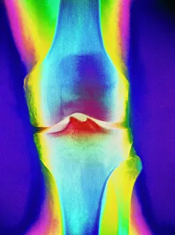
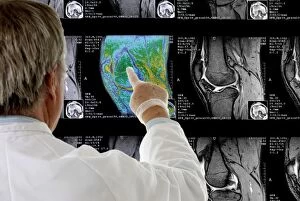
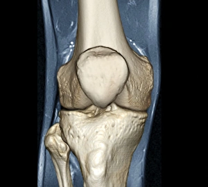
The patella, also known as the kneecap, is a vital component of the human knee joint and can be seen clearly in a coloured X-ray of a human knee joint


All Professionally Made to Order for Quick Shipping
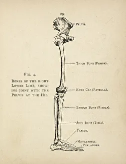
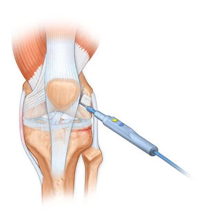
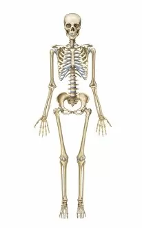
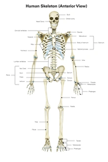
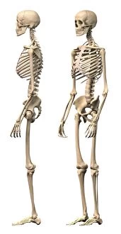
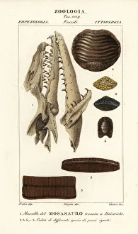
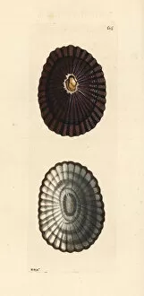
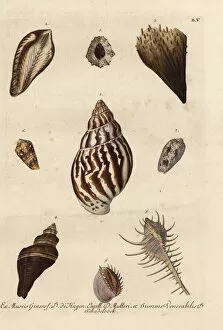
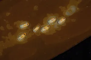
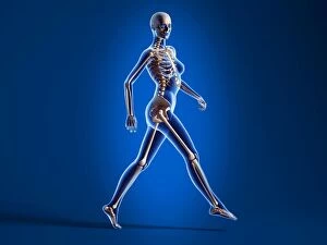
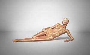
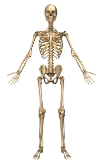
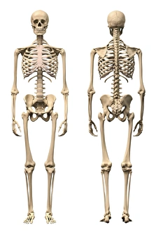
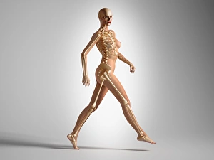
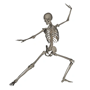
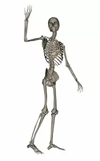
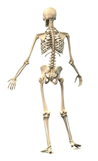
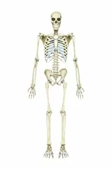
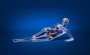
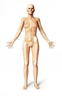
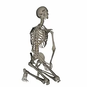
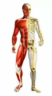
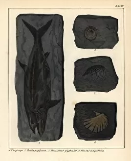
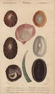
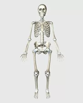
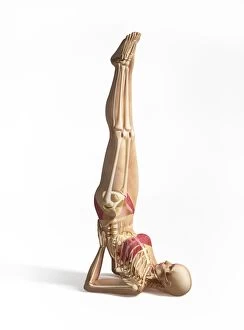
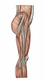
The patella, also known as the kneecap, is a vital component of the human knee joint and can be seen clearly in a coloured X-ray of a human knee joint, standing out amidst the surrounding bones. In a diagram depicting the bones of the right leg and hip, the patella is highlighted as an essential part of this intricate system. When it comes to medical procedures involving the patella, precision is key. A bovie instrument is used to cut through retincaculum and clean up femur in cases where there is displacement or damage to this crucial bone within the knee joint. An illustration showcasing the anterior view of a knee provides insight into its articular surface meniscus. This detailed depiction allows us to understand how these structures work together for optimal movement and stability. Interestingly enough, even nature has its own version of "patella. " In United Kingdom's Scotland region lies Moray's Lossiemouth - home to a vibrant limpet colony on rocks. The blue-rayed limpet (Patella pellucida) thrives here, showcasing its unique adaptation that resembles our very own kneecap. Zooming out from specific anatomy studies, we can appreciate how integral the patella is within our entire skeletal system. Whether viewed from front or side angles with labels or depicted in perspective view alongside other male skeleton parts – it becomes evident that every bone plays an important role in maintaining our body's structure and function. To further highlight diversity across species, one might explore fossils like Mososaurus skull and fish palates. These remnants remind us that while humans have evolved complex anatomical systems such as our knees' intricate design including patellas – other creatures have their own fascinating adaptations too. Whether we examine it through medical images or delve into natural wonders like limpets or fossils – exploring various aspects related to "patella" brings about awe for both human anatomy and biodiversity.