
Photosynthetic Collection
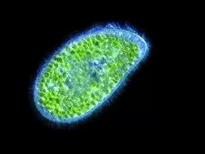
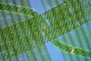
Photosynthetic organisms, such as diatoms, are fascinating microscopic creatures that play a crucial role in our ecosystem


All Professionally Made to Order for Quick Shipping
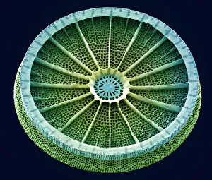
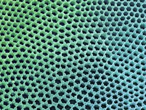
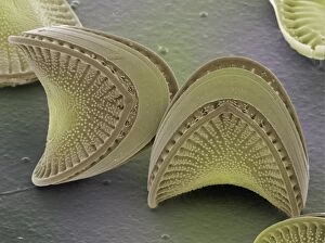
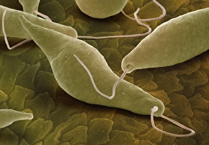
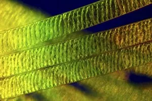
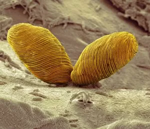
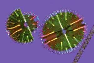
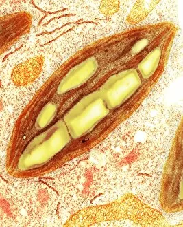
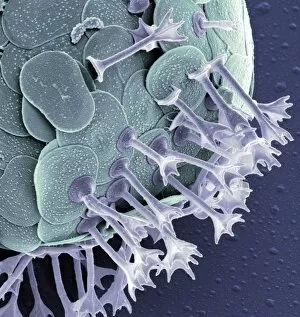
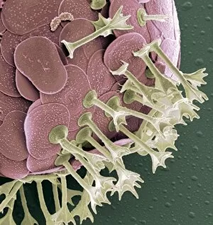
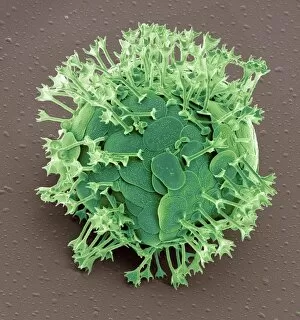
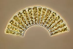
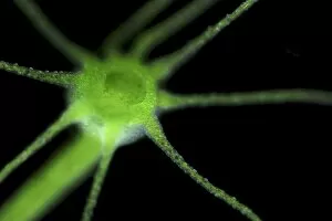
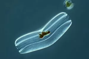
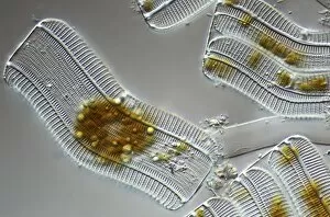
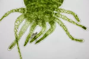
Photosynthetic organisms, such as diatoms, are fascinating microscopic creatures that play a crucial role in our ecosystem. Seen here under the scanning electron microscope (SEM), their intricate structures and vibrant colors captivate scientists and artists alike. One of the most striking features of diatoms is their cell wall, which can be observed in detail through SEM imaging. These walls are made up of silica, forming beautiful geometric patterns that resemble delicate artwork. Each diatom species has its own unique design, making them a treasure trove for researchers studying biodiversity. Beyond their aesthetic appeal, diatoms possess chloroplasts - specialized organelles responsible for photosynthesis. These tiny powerhouses convert sunlight into energy by harnessing pigments like chlorophyll. Under SEM magnification, we can witness the complexity of these chloroplast structures within diatom cells. Diatoms come in various shapes and sizes; some appear like elongated rods while others resemble intricate snowflakes or even miniature fans. SEM allows us to explore this diversity up close and appreciate the incredible intricacy present within each individual organism. These photosynthetic wonders not only contribute to global oxygen production but also serve as an essential food source for marine life. Their ability to thrive in diverse environments makes them vital indicators of water quality and ecological health. Through continued research using techniques like SEM imaging, we gain valuable insights into the world organisms like diatoms. By understanding their structure and function at a microscopic level, we unlock secrets about our planet's past climate conditions while also paving the way for innovative solutions inspired by nature's ingenuity.