Fraud Blocker Skip to main content

Plankton Collection (page 6)

Plankton: Unveiling the Hidden Marvels of the Ocean Dive into the mesmerizing world of plankton, where beauty and wonder intertwine in a delicate dance

Background imagePlankton Collection: Microalgae, light micrograph

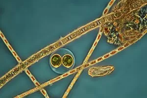
Microalgae, light micrograph
Microalgae. Differential interference contrast (DIC) micrograph of Botryococcus braunii (round), and other species of green alga. Magnification: x278, when printed 10 centimetres wide

Background imagePlankton Collection: Haematococcus algae, light micrograph

Haematococcus algae, light micrograph
Haematococcus algae. Differential interference contrast (DIC) micrograph of a colony of Haematococcus sp. green alga. Magnification: x220, when printed 10 centimetres wide

Background imagePlankton Collection: Rotifer, light micrograph

Rotifer, light micrograph
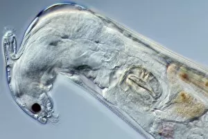
Rotifer. Darkfield illuminated light micrograph of a rotifer (phylum Rotifera). Rotifers (phylum Rotifera) are microscopic aquatic animals that are related to roundworms

Background imagePlankton Collection: Fossil foraminifera necklace C016 / 5995

Fossil foraminifera necklace C016 / 5995
Fossil foraminifera (Alveolina sp.) necklace from Sindh, India. The fossils are from Eocene limestone. Necklace from John Whittakers cabinet. Natural History Museum, London, UK

Background imagePlankton Collection: Foraminiferan shell, SEM C018 / 0312

Foraminiferan shell, SEM C018 / 0312
Foraminiferan shell, SEM

Background imagePlankton Collection: Scenedesmus algae, SEM C014 / 1462

Scenedesmus algae, SEM C014 / 1462
Scenedesmus algae. Coloured scanning electron micrograph (SEM) of Scenedesmus sp. algae. Scenedesmus is a nonmotile colonial green algae that has its cells arranged in a row

Background imagePlankton Collection: Diatom frustules (SEM) C014 / 2412

Diatom frustules (SEM) C014 / 2412
Diatom frustules. Coloured scanning electron micrograph of diatom frustules (Cymbella gastroides). Diatoms are a group of photosynthetic, single-celled algae containing about 10, 000 species

Background imagePlankton Collection: Diatom frustules (SEM) C014 / 2147

Diatom frustules (SEM) C014 / 2147
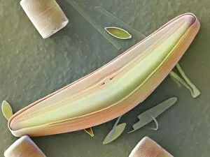
Diatom frustules. Coloured scanning electron micrograph of the internal surface of a diatom frustule of Cymbella gastroides. The tiny diatom inside the cymbella is navicula sp
Background imagePlankton Collection: Diatoms, light micrograph C014 / 4675

Diatoms, light micrograph C014 / 4675
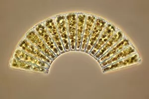
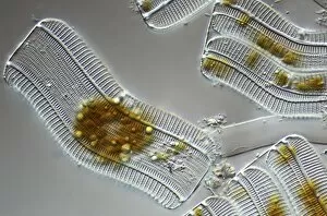
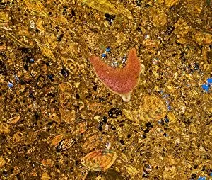
Diatoms. Differential interference contrast micrograph of a group of Meridion circulare diatoms. Diatoms are a group of photosynthetic, single-celled algae containing about 10, 000 species

Background imagePlankton Collection: Diatom, light micrograph C014 / 4677

Diatom, light micrograph C014 / 4677
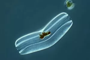
Diatom. Differential interference contrast micrograph of a Campylodiscus sp. diatom. Diatoms are a group of photosynthetic, single-celled algae containing about 10, 000 species

Background imagePlankton Collection: Green algae, light micrograph C014 / 4670

Green algae, light micrograph C014 / 4670
Green algae. Differential interference contrast micrograph of a single green alga, showing the chloroplast (green) inside. Algae use the chloroplasts to produce food through photosynthesis

Background imagePlankton Collection: Diatoms, light micrograph C014 / 4671

Diatoms, light micrograph C014 / 4671
Diatoms. Differential interference contrast micrograph of a group of Achnantes longipes diatoms. Diatoms are a group of photosynthetic, single-celled algae containing about 10, 000 species

Background imagePlankton Collection: Anabaena algae, SEM

Anabaena algae, SEM
Anabaena algae. Coloured scanning electron micrograph (SEM) of Anabaena sp. freshwater alga from a pond. Anabaena is a filamentous species of blue-green algae (cyanobacteria)

Background imagePlankton Collection: Radiolarian, SEM C014 / 4862

Radiolarian, SEM C014 / 4862
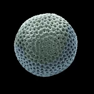
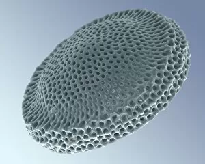
Radiolarian. Coloured scanning electron micrograph (SEM) of the shell or test of a radiolarian. Radiolaria are single-celled protozoans that are found in marine plankton

Background imagePlankton Collection: Diatom, light micrograph C014 / 4669

Diatom, light micrograph C014 / 4669
Diatom. Differential interference contrast micrograph of an Achnantes longipes diatom. Diatoms are a group of photosynthetic, single-celled algae containing about 10, 000 species

Background imagePlankton Collection: Radiolarian, SEM C014 / 4863

Radiolarian, SEM C014 / 4863
Radiolarian. Coloured scanning electron micrograph (SEM) of the shell or test of a radiolarian. Radiolaria are single-celled protozoans that are found in marine plankton

Background imagePlankton Collection: Diatoms, light micrograph C014 / 4668

Diatoms, light micrograph C014 / 4668
Diatoms. Differential interference contrast micrograph of a group of Achnantes longipes diatoms. Diatoms are a group of photosynthetic, single-celled algae containing about 10, 000 species

Background imagePlankton Collection: Diatoms, light micrograph C014 / 4674

Diatoms, light micrograph C014 / 4674
Diatoms. Differential interference contrast micrograph of a group of Achnantes longipes diatoms. Diatoms are a group of photosynthetic, single-celled algae containing about 10, 000 species

Background imagePlankton Collection: Rotifer, light micrograph C014 / 4666

Rotifer, light micrograph C014 / 4666
Rotifer, differential interference contrast micrograph. These animals are related to roundworms. They are usually freshwater organisms though a few marine sp

Background imagePlankton Collection: Bdelloid rotifer, SEM

Bdelloid rotifer, SEM
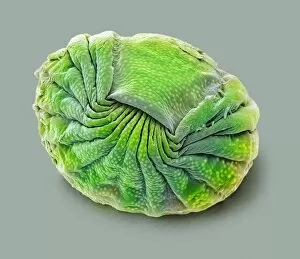
Bdelloid rotifer. Coloured scanning electron micrograph (SEM) of a rotifer (class Bdelloidea) from a pond. Rotifers (phylum Rotifera) are microscopic aquatic animals that are related to roundworms

Background imagePlankton Collection: Foraminiferan

Foraminiferan, darkfield light micrograph. Foraminifera are marine single-celled protozoa that construct and inhabit shells composed of several chambers

Background imagePlankton Collection: Diatom, light micrograph C016 / 8606

Diatom, light micrograph C016 / 8606
Diatom. Phase-contrast light micrograph of a diatom from the Mediterranean. Diatoms are a group of photosynthetic, single-celled algae containing about 10, 000 species

Background imagePlankton Collection: Diatoms, light micrograph C016 / 8605

Diatoms, light micrograph C016 / 8605
Diatoms, Shearing contrast light micrograph. Diatoms are a group of photosynthetic, single-celled algae containing about 10, 000 species

Background imagePlankton Collection: Diatoms, light micrograph C016 / 8598

Diatoms, light micrograph C016 / 8598
Diatoms. Interphase contrast light micrograph of a selection of differently shaped diatoms. Diatoms are a group of photosynthetic, single-celled algae containing about 10, 000 species

Background imagePlankton Collection: Diatoms, light micrograph C016 / 8602

Diatoms, light micrograph C016 / 8602
Diatoms. Phase-contrast light micrograph of diatoms from the Mediterranean. Diatoms are a group of photosynthetic, single-celled algae containing about 10, 000 species

Background imagePlankton Collection: Foraminifera, light micrograph C016 / 8597

Foraminifera, light micrograph C016 / 8597
Foraminifera. Interphase contrast light micrograph of a selection of different foraminifera. Foraminifera are marine single-celled protozoa that construct

Background imagePlankton Collection: Diatoms, light micrograph C016 / 8600

Diatoms, light micrograph C016 / 8600
Diatoms. Differential interference contrast (DIC) light micrograph of diatoms from the Mediterranean. Diatoms are a group of photosynthetic, single-celled algae containing about 10, 000 species

Background imagePlankton Collection: Green algae, light micrograph C016 / 8595

Green algae, light micrograph C016 / 8595
Green algae, Interphase contrast light micrograph. Magnification: x280, when printed 10 centimetres wide

Background imagePlankton Collection: Fossil diatom, light micrograph C016 / 8586

Fossil diatom, light micrograph C016 / 8586
Fossil diatom. Interphase contrast light micrograph of a fossilised diatom. Diatoms are a group of photosynthetic, single-celled algae containing about 10, 000 species

Background imagePlankton Collection: Rotifer with eggs, light micrograph C016 / 8588

Rotifer with eggs, light micrograph C016 / 8588
Rotifer with eggs. Differential interference contrast (DIC) light micrograph of a rotifer (phylum Rotifera) carrying eggs (small, round)

Background imagePlankton Collection: Diatoms, light micrograph C016 / 8585

Diatoms, light micrograph C016 / 8585
Diatoms. Interphase contrast light micrograph of two differently shaped diatoms. Diatoms are a group of photosynthetic, single-celled algae containing about 10, 000 species

Background imagePlankton Collection: Fossil diatom, light micrograph C016 / 8583

Fossil diatom, light micrograph C016 / 8583
Fossil diatom. Interphase contrast light micrograph of a fossilised diatom. Diatoms are a group of photosynthetic, single-celled algae containing about 10, 000 species

Background imagePlankton Collection: Copepod larva, polarised light micrograph C016 / 8577

Copepod larva, polarised light micrograph C016 / 8577
Copepod larva. Polarised light micrograph of a nauplius larva. A nauplius is the first larval stage of a crustacean in which the thorax and abdomen have not developed yet

Background imagePlankton Collection: Microalgae research

Microalgae research. Biologist holding a Petri dish containing cultured diatom microalgae. Diatoms are photosynthetic, single-celled algae that form an important part of the plankton at the base of

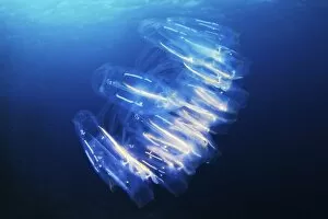
Background imagePlankton Collection: Salp colony

Salp colony (Cyclosalpa). Salps are tubular shaped gelatinous marine animals. Multiple individuals join together to form long colonies that feed on phytoplankton

Background imagePlankton Collection: Amphipod crustacean

Amphipod crustacean. Close-up of the amphipod (Anonyx nugax). Amphipods typically have a laterally flattened body and fourteen limbs

Background imagePlankton Collection: Fossil-bearing rock, polarised LM C017 / 8483

Fossil-bearing rock, polarised LM C017 / 8483
Fossil-bearing rock, polarised light micrograph. This thin slice from a limestone rock shows various Nummulite fossils. It is a type known as Nummulitie limestone

Background imagePlankton Collection: Fossilised foraminiferan, SEM C015 / 5772

Fossilised foraminiferan, SEM C015 / 5772
Fossilised foraminiferan. Scanning electron micrograph (SEM) of a section through the fossilised shell of a foraminiferan in a sample of limestone

Background imagePlankton Collection: Formation of fossil fuels, artwork C017 / 8034

Formation of fossil fuels, artwork C017 / 8034
Formation of fossil fuels. Computer artwork showing how fossil fuels, such as coil, oil and gas, are formed. Fossil fuels are rich in hydrocarbons

Background imagePlankton Collection: Golden algae, light micrograph

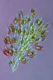
Golden algae, light micrograph
Golden algae. Polarised light micrograph of a colony of Dinobryon sp. golden algae (chrysophytes). Magnification: x200 when printed 10 centimetres wide

Background imagePlankton Collection: William Carpenter, British naturalist C016 / 5058

William Carpenter, British naturalist C016 / 5058
William Benjamin Carpenter (1813-1885), British naturalist. Carpenter trained as a physician, but published widely in many fields

Background imagePlankton Collection: Didymograptus, graptolite fossil C016 / 4848

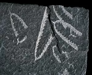
Didymograptus, graptolite fossil C016 / 4848
Didymograptus, graptolite fossil. Graptolites are an extinct group of marine, colonial animals. Known for their tuning-fork shape, they formed part of ancient plankton

Background imagePlankton Collection: Indian Ocean, chlorophyll concentration C016 / 3722

Indian Ocean, chlorophyll concentration C016 / 3722
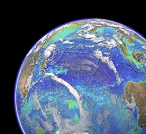
Indian Ocean, chlorophyll concentration and clouds. Computer graphic of the Indian Ocean on an Earth globe, generated using satellite and mapping data

Background imagePlankton Collection: Indian Ocean, chlorophyll and bathymetry C016 / 3721

Indian Ocean, chlorophyll and bathymetry C016 / 3721
Indian Ocean, chlorophyll concentration and bathymetry with clouds. Computer graphic of the Indian Ocean on an Earth globe, generated using satellite and mapping data

Background imagePlankton Collection: Indian Ocean, chlorophyll and bathymetry

Indian Ocean, chlorophyll and bathymetry
Indian Ocean, chlorophyll concentration and bathymetry. Computer graphic of the Indian Ocean on an Earth globe, generated using satellite and mapping data


All Professionally Made to Order for Quick Shipping

Plankton: Unveiling the Hidden Marvels of the Ocean Dive into the mesmerizing world of plankton, where beauty and wonder intertwine in a delicate dance. From the intricate artistry of diatom algae to the awe-inspiring sight of a whale shark feeding with its mouth wide open, these tiny organisms hold immense significance in our vast oceans. Ernst Haeckel's stunning depiction of diatom algae showcases their exquisite patterns and shapes, reminding us that even microscopic life forms can be true works of art. These diatoms play a crucial role as primary producers, contributing to almost half of Earth's oxygen production. Imagine being a diver off Australia's coast, witnessing firsthand the majestic encounter between a whale shark and its planktonic feast. This gentle giant glides through the water effortlessly, creating an ethereal spectacle that captivates divers from around the world. Examining diatoms under scanning electron microscopy reveals their intricate structures up close. Each detail unravels another layer of complexity within these minuscule organisms, highlighting their importance as essential components in marine ecosystems. The Isle of Man becomes home to basking sharks like Certorhinus maximus during certain times of year. Douglas David Seifert's photograph captures one such moment when this magnificent creature graces us with its presence. Its sheer size reminds us how vital plankton is for sustaining these gentle giants' colossal appetite. In every drop sampled from marine environments lies an entire universe waiting to be explored - each diatom telling its unique story. Through SEM imagery, we gain insight into their diverse forms and functions; they are truly nature's architects shaping our oceans' delicate balance. Returning once again to Cenderawasih Bay in West Papua Indonesia brings yet another breathtaking encounter with whale sharks – Rhincodon typus – gracefully swimming amidst clouds of planktonic abundance. Their presence serves as a reminder that protecting these fragile ecosystems is crucial for the survival of these magnificent creatures.