Fraud Blocker Skip to main content

Plant Anatomy Collection (page 3)

"Exploring the Intricate World of Plant Anatomy: A Fascinating Journey through Micrographs and Artwork" Delve into the hidden wonders with these captivating images

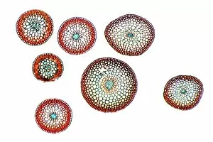
Background imagePlant Anatomy Collection: Moss stems, light micrograph

Moss stems, light micrograph. Transverse section through stems of a cord moss (Polytrichum commune). The outside of the stems, the epidermis, is made up of thick-walled cells

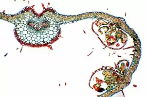
Background imagePlant Anatomy Collection: Fern frond, light micrograph

Fern frond, light micrograph. Transverse section through the frond of a fern (Cyrtomium falcatum), showing the sporangia. At left is a central vascular bundle (meristele)

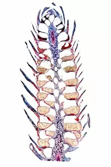
Background imagePlant Anatomy Collection: Clubmoss cone, light micrograph

Clubmoss cone, light micrograph. Longitudinal section through the cone (strobilus) of the clubmoss Lycopodium clavatum. The strobilus consists of a central axis (purple)

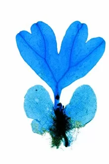
Background imagePlant Anatomy Collection: Fern plant reproduction, light micrograph

Fern plant reproduction, light micrograph. At bottom, the double kidney-shaped gametophyte (prothallus) plant has anchored itself with root-like rhizoids (central black area)

Background imagePlant Anatomy Collection: Fern stem, light micrograph

Fern stem, light micrograph. Transverse section through a rachis (stem) of the bracken fern (Pteridium aquilinum). Under the outer epidermis (black) is a thin cortex (deep red)

Background imagePlant Anatomy Collection: Horsetail cone, light micrograph

Horsetail cone, light micrograph. Longitudinal section through a young cone (strobilus) of the rough horsetail (Equisetum hyemale)

Background imagePlant Anatomy Collection: Liverwort spore capsule, light micrograph

Liverwort spore capsule, light micrograph. Longitudinal section through the thallus and sporangium of a liverwort (Jungammania sp.) The sporangium (spore capsule)

Background imagePlant Anatomy Collection: Moss reproductive parts, light micrograph

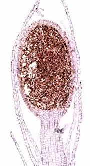
Moss reproductive parts, light micrograph. Longitudinal section through the antheridial cup (male reproductive parts) of a cord moss (Polytrichum commure)

Background imagePlant Anatomy Collection: Liverwort spore elaters, light micrograph

Liverwort spore elaters, light micrograph. Transverse section through the sporangium of a liverwort (Pellia epiphylla). This is the basal portion of the sporangium

Background imagePlant Anatomy Collection: Moss spore capsule, light micrograph

Moss spore capsule, light micrograph
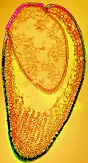
Moss spore capsule, polarised light micrograph. Longitudinal section through a spore capsule from a fire moss (Funaria hygrometrica)

Background imagePlant Anatomy Collection: Horsetail stem, light micrograph

Horsetail stem, light micrograph. Transverse section through the stem of the common horsetail (Equisetum arvensis). The stem consists of an outer epidermis and an outer cortex of sclerenchyma cells

Background imagePlant Anatomy Collection: Mushroom gill cap, light micrograph

Mushroom gill cap, light micrograph. Transverse section through the cap of a common mushroom (Psalliota sp.) showing the gill lamellae (white)

Background imagePlant Anatomy Collection: Fern rhizome, light micrograph

Fern rhizome, light micrograph. Transverse section through the center of a rhizome from the Killarney fern (Trichomanes speciosum)

Background imagePlant Anatomy Collection: Liverwort spores, light micrograph

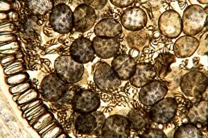
Liverwort spores, light micrograph. Transverse section through the sporangium of a liverwort (Pellia epiphylla). Part of the sporangiums outer wall is at left

Background imagePlant Anatomy Collection: Clubmoss stem, light micrograph

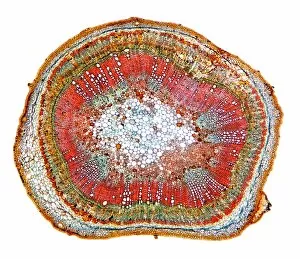
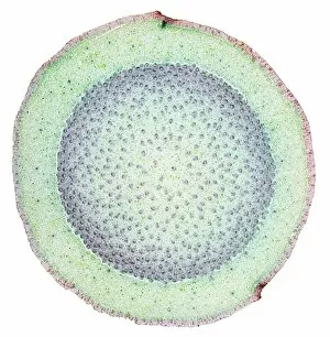
Clubmoss stem, light micrograph
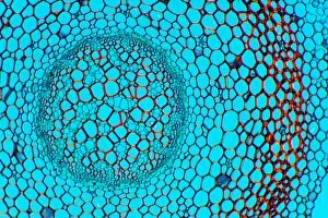
Clubmoss stem, polarised light micrograph. Transverse section through the stem of the clubmoss Lycopodium clavatum. This is the central portion of the stem consisting of the inner cortex (red)

Background imagePlant Anatomy Collection: Rust fungus infection, light micrograph

Rust fungus infection, light micrograph. Transverse section through a barberry (Berberis sp.) leaf infected by a parasitic rust (Puccinia sp.)

Background imagePlant Anatomy Collection: Fern spore capsule, light micrograph

Fern spore capsule, light micrograph
Fern spore capsule, polarised light micrograph. Transverse section through the spororocarp (spore capsule) of the water fern pillwort (Pilularia globulifera)

Background imagePlant Anatomy Collection: Mould fruiting bodies, light micrograph

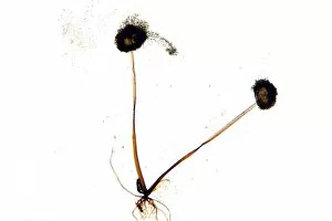
Mould fruiting bodies, light micrograph. Black mould (Rhizopus nigricans) fruiting bodies (top and upper right) liberating spores

Background imagePlant Anatomy Collection: Algal colony, light micrograph

Algal colony, light micrograph. Colony of the green freshwater alga Coleochaete sentata. The whole plant is a disc composed of filaments packed close together, branching out from the centre

Background imagePlant Anatomy Collection: Leaf section, SEM

Leaf section, SEM
Leaf section. Coloured scanning electron micrograph (SEM) of a section through a fractured leaf. At top is a single layer of cells that forms the epidermis of the leaf

Background imagePlant Anatomy Collection: Lily bud, light micrograph

Lily bud, light micrograph
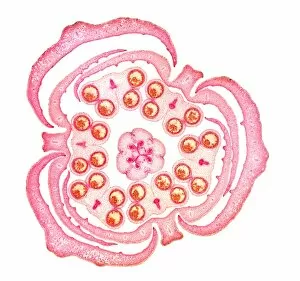
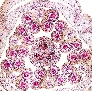
Lily bud. Light micrograph of a cross-section through an immature lily bud (Lilium sp.), showing the male and female reproductive organs of the flower

Background imagePlant Anatomy Collection: Maize niblet, light micrograph

Maize niblet, light micrograph
Maize niblet. Light micrograph of a maize niblet, the seed of the maize plant (Zea mays). A seed contains an embryo, a fertilised ovule from which a new plant can grow (green, upper left)

Background imagePlant Anatomy Collection: Butchers broom stem, light micrograph

Butchers broom stem, light micrograph
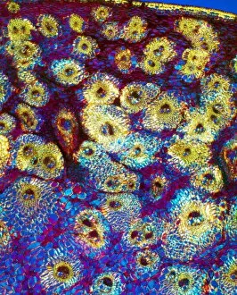
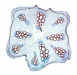
Butchers broom stem. Polarised light micrograph of a section through a butchers broom stem (Ruscus aculeatis) showing many vascular bundles (dark clusters) within the plant cortex (orange)

Background imagePlant Anatomy Collection: Umbrella grass stem, light micrograph

Umbrella grass stem, light micrograph
Umbrella grass stem. Polarised light micrograph of a section through a umbrella grass stem (Cyperus alternifolius). Vascular bundles (brown) can be seen containing xylem (larger openings)

Background imagePlant Anatomy Collection: Flax plant stem, light micrograph

Flax plant stem, light micrograph
Flax plant stem. Light micrograph of a transverse section through a stem of the flax plant (Linum usitatissimum). The layers from outer to inner (some very thin) are the epidermis (bottom)

Background imagePlant Anatomy Collection: Purple moor grass stem, light micrograph

Purple moor grass stem, light micrograph
Purple moor grass stem. Polarised light micrograph of a section through a purple moor grass stem (Molinia caerula). Vascular bundles (round orange structures)

Background imagePlant Anatomy Collection: Clover plant stem, light micrograph

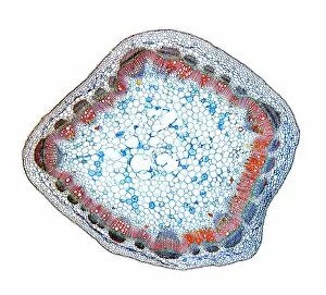
Clover plant stem, light micrograph
Clover plant stem. Light micrograph of a transverse section through a stem of the clover plant (Trifolium pratense). Magnification: x100 when printed at 10 centimetres wide

Background imagePlant Anatomy Collection: Bamboo stem, light micrograph

Bamboo stem, light micrograph
Bamboo stem. Polarised light micrograph of a section through a bamboo stem (Bambusa vulgaris). Vascular bundles can be seen (yellow/green) containing xylem (larger openings)

Background imagePlant Anatomy Collection: Maize stem, light micrograph

Maize stem, light micrograph
Maize stem. Polarised light micrograph of a section through a maize plant stem (Zea mays). A vascular bundles (brown) can be seen containing xylem (larger openings)

Background imagePlant Anatomy Collection: Spiny rush stem, light micrograph

Spiny rush stem, light micrograph
Spiny rush stem. Polarised light micrograph of a section through a spiny rush stem (Juncus actus). Vascular bundles can be seen (purple/red) containing xylem (larger openings)

Background imagePlant Anatomy Collection: Wheat stem, light micrograph

Wheat stem, light micrograph
Wheat stem. Light micrograph of a section through the stem of a wheat grass (Triticum aestivium). The circular structures (orange and green) are vascular bundles

Background imagePlant Anatomy Collection: Sugar cane stem, light micrograph

Sugar cane stem, light micrograph
Sugar cane stem. Polarised light micrograph of a section through a sugar cane stem (Saccharum officinareum). Vascular bundles (round blue/purple structures)

Background imagePlant Anatomy Collection: Lily anther, polar light micrograph

Lily anther, polar light micrograph
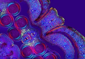
Lily anther. Polar light micrograph of a cross-section through a lily flower anther (Lilium sp.), showing four anther sacs (round structures)

Background imagePlant Anatomy Collection: Reed canary grass stem, light micrograph

Reed canary grass stem, light micrograph
Reed canary grass stem. Light micrograph of a section through a reed canary grass stem (Phalaris arundinacea). Vascular bundles (oval yellow and red structures)

Background imagePlant Anatomy Collection: Cotton plant root, light micrograph

Cotton plant root, light micrograph
Cotton plant root. Light micrograph of a transverse section through a root of the cotton plant (Gossypium hirsutum). The layers from outer to centre (some very thin)

Background imagePlant Anatomy Collection: Dragon tree stem, light micrograph

Dragon tree stem, light micrograph
Dragon tree stem. Light micrograph of a section through a dragon tree stem (Dracaena draco). Vascular bundles (round dark structures) can be seen containing xylem (larger openings)

Background imagePlant Anatomy Collection: Peanut plant stem, light micrograph

Peanut plant stem, light micrograph
Peanut plant stem. Light micrograph of a transverse section through a stem of the peanut plant (Arachis hypogaea). Below the stems outer layer (epidermis) is a cortex of parenchyma cells (blue)

Background imagePlant Anatomy Collection: Lily anther, light micrograph

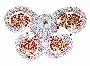
Lily anther, light micrograph
Lily anther. Light micrograph of a cross-section through a lily flower anther (Lilium sp.), showing four anther sacs (curled structures)

Background imagePlant Anatomy Collection: Cucurbita plant stem, light micrograph

Cucurbita plant stem, light micrograph
Cucurbita plant stem. Light micrograph of a transverse section through a stem of a Cucurbita plant. Magnification: x14 when printed at 10 centimetres wide

Background imagePlant Anatomy Collection: Lily carpel, LM

Lily carpel, LM
Lily carpel. Light micrograph of a cross-section through a lily flower carpel (Lilium sp.), showing the three fused leaves that make up the pistil

Background imagePlant Anatomy Collection: Lily bud pistil and anthers, LM

Lily bud pistil and anthers, LM
Lily bud. Light micrograph of a cross-section through an immature lily bud (Lilium sp.), showing the male and female reproductive organs of the flower


All Professionally Made to Order for Quick Shipping

"Exploring the Intricate World of Plant Anatomy: A Fascinating Journey through Micrographs and Artwork" Delve into the hidden wonders with these captivating images. From dicotyledon plant stems to castor oil stems, each light micrograph unveils intricate structures that support life. Witness the mesmerizing beauty of Rhynchoglossum obliquum in artwork C016 / 5646, as its delicate details come to life. Dive beneath the surface with a glimpse at maize roots captured under a light microscope. Marvel at the complexity of English oak leaf pores revealed through scanning electron microscopy (SEM), showcasing nature's precision engineering. Take a closer look at the enchanting buttercup flower and tomato leaf, both magnified by light micrography. Explore pollination like never before, as SEM captures this pivotal moment in stunning detail. Uncover secrets hidden within French lavender leaf pores using SEM, revealing their unique structure and function. Witness gutation water droplets on dicot plants' leaf hairs through an unknown magnification SEM image - a phenomenon that showcases nature's ability to adapt and thrive. Embark on an exploration of Ladys mantle reproductive parts with SEM imagery, offering insights into reproduction strategies employed by these remarkable plants. Finally, get up close and personal with primrose pollen under SEM - tiny particles that play a crucial role in plant reproduction. Join us on this visual journey where science meets artistry; discover how every nook and cranny holds mysteries waiting to be unraveled in the fascinating world of plant anatomy.