
Podocyte Collection
The podocyte, a crucial component of the kidney's filtration system, plays a vital role in maintaining our body's fluid balance


All Professionally Made to Order for Quick Shipping
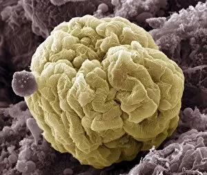
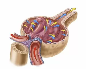
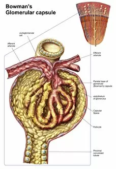
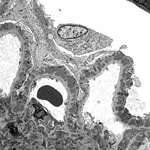
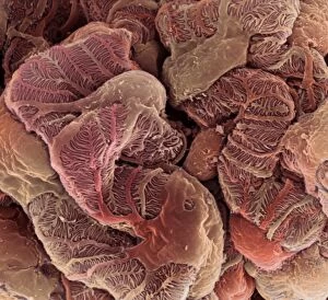
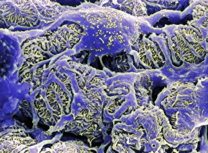
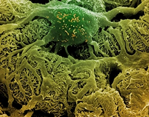
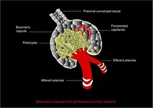
The podocyte, a crucial component of the kidney's filtration system, plays a vital role in maintaining our body's fluid balance. Found within the renal glomerulus, these specialized cells form intricate foot-like extensions that wrap around capillaries, creating a unique structure known as the filtration membrane. Through their delicate interdigitating processes called pedicels, podocytes create tiny gaps or slits called filtration slits. These slits allow for selective passage of essential substances while preventing larger molecules like proteins from escaping into the urine. In an artist's depiction of the glomerulus capillaries, we can visualize how podocytes embrace and support these blood vessels with their complex network of foot processes. This arrangement ensures efficient filtration by providing both structural stability and flexibility to adapt to varying pressures. When examining the anatomy of Bowman's glomerular capsule under a scanning electron microscope (SEM), we witness the intricate architecture formed by podocytes surrounding it. Their presence is crucial in maintaining proper function and preventing leakage during ultrafiltration. Further magnification using transmission electron microscopy (TEM) reveals detailed images showcasing individual podocytes' fine structures within the glomerulus. The TEM images provide us with invaluable insights into their morphology and organization at an even more microscopic level. These captivating SEM images capture multiple views of kidney glomeruli, emphasizing how integral they are to this complex organ's functionality. Their strategic placement allows them to regulate fluid balance effectively while ensuring waste products are eliminated efficiently through urine formation. In summary, whether observed through SEM or TEM imaging techniques or depicted by talented artists capturing its essence visually, exploring the world of podocytes offers us glimpses into one aspect of renal physiology that keeps our bodies functioning optimally – filtering out waste while preserving essential substances for overall health and well-being.