
Pollen Grains Collection
"Pollen Grains: Nature's Tiny Messengers of Life" Pollen grains, the microscopic wonders of the plant kingdom


All Professionally Made to Order for Quick Shipping
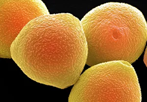
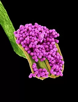
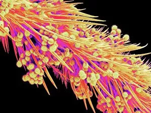
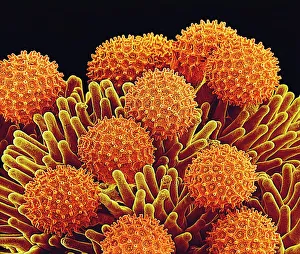
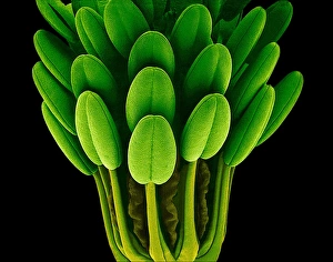
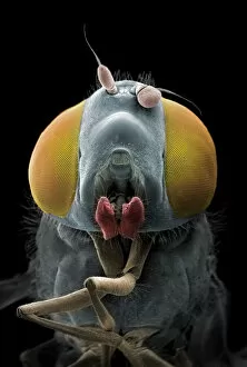
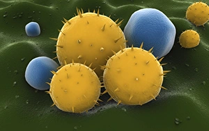
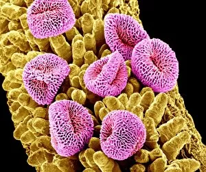
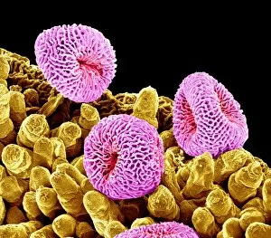
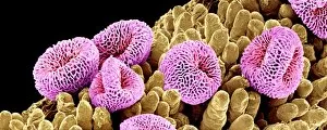
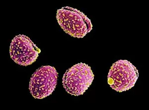
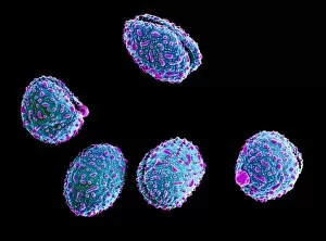
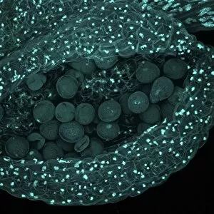
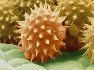
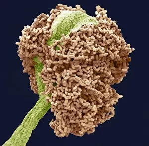
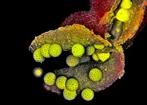
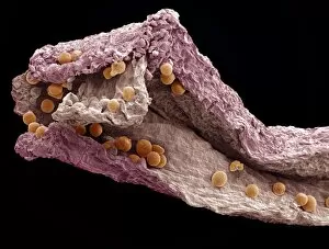
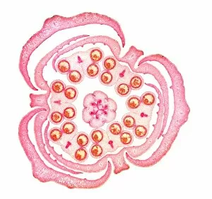
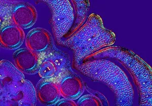
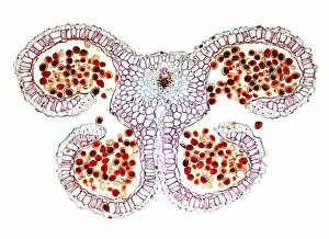
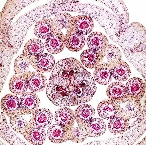
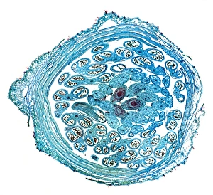
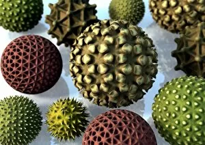
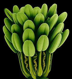
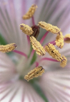
"Pollen Grains: Nature's Tiny Messengers of Life" Pollen grains, the microscopic wonders of the plant kingdom, play a crucial role in the reproduction and survival of countless plant species. Through their intricate structures and diverse appearances, these tiny particles hold fascinating secrets waiting to be explored. Under the scanning electron microscope (SEM), we delve into a world unseen by the naked eye. The Geranium anther reveals its delicate beauty as it releases pollen grains with precision and grace. Similarly, the Honeybee leg showcases its remarkable ability to collect and transport these vital particles from flower to flower. Moving on to Tea flower stamens captured under SEM, we witness nature's artistry at its finest. Each filament carries numerous pollen grains like precious gems adorning a crown. Meanwhile, Geranium pollen takes center stage with its distinct shape and texture – a testament to evolution's ingenuity. Hazel pollen grains come into focus next; their unique design allows them to withstand harsh conditions during wind pollination. Morning glory pollen exhibits mesmerizing patterns that captivate our imagination while Columbine flower stamens showcase an array of colors fit for an artist's palette. The Bay tree anther stands out with its intricate network of chambers housing countless pollen grains ready for dispersal. Common daisy anthers reveal their vibrant yellow hue as they burst open, releasing clouds of life-giving dust into the air. Euphorbia flower reproductive parts offer us a glimpse into nature's complexity – each component working harmoniously towards successful pollination. Finally, Geranium pollen once again graces our view under SEM; this time revealing minute details that make it perfectly adapted for cross-pollination. In this captivating journey through SEM images, we gain appreciation for these minuscule yet mighty entities known as "pollen grains. " They are not merely specks floating in the breeze but rather essential messengers ensuring biodiversity thrives across our planet.