
Polysaccharide Collection
"Exploring the Fascinating World of Polysaccharides: From Cellulose Nanorods to Intestinal Microvilli" In this captivating conceptual image


All Professionally Made to Order for Quick Shipping
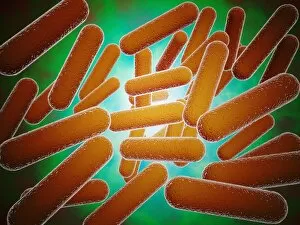
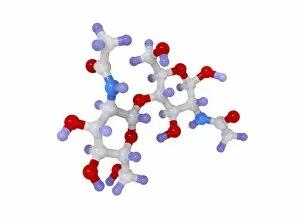
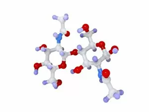
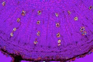
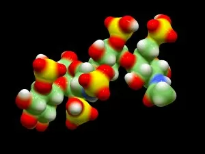
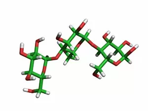
"Exploring the Fascinating World of Polysaccharides: From Cellulose Nanorods to Intestinal Microvilli" In this captivating conceptual image, we get a glimpse into the intricate structure of cellulose nanorods. These tiny rods, composed of polysaccharides, play a crucial role in providing strength and rigidity to plant cell walls. Moving on to another mesmerizing SEM image, we witness the delicate beauty of intestinal microvilli. These finger-like projections are covered with a layer of polysaccharides that aid in nutrient absorption within our digestive system. Shifting our focus to chitin, we encounter an enchanting molecular model. This polysaccharide is found abundantly in nature and forms the exoskeletons of insects and crustaceans. Its unique properties make it an essential component for various applications ranging from biomedical materials to environmental solutions. Now let's delve into the fascinating world beneath the surface as we explore a light micrograph showcasing Dahlia tubers. The intricate network seen here is made up of complex carbohydrates known as polysaccharides that provide energy storage for plants during dormancy periods. Zooming out from individual cells, we take a look at the bigger picture – plant cell walls depicted in a detailed diagram. Composed primarily of cellulose along with other polysaccharides like hemicellulose and pectin, these structures provide support and protection while allowing for flexibility and growth. Last but not least, behold the artistic representation of glycogen molecules – highly branched chains formed by glucose units linked together through glycosidic bonds. As one of nature's primary energy storage compounds found mainly in animals' liver and muscles, glycogen plays a vital role in maintaining blood sugar levels during fasting or exercise. From cellulose nanorods shaping plant architecture to chitin fortifying insect armor.