
Prokaryotic Collection
"Exploring the Intricate World Cells: A Fusion of Science and Art" Step into a mesmerizing realm where science meets art, as we delve into the captivating world cells


All Professionally Made to Order for Quick Shipping
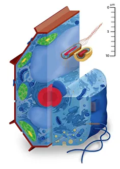
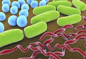
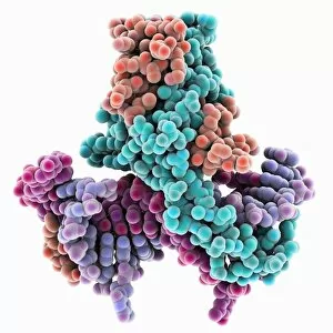
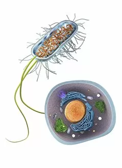
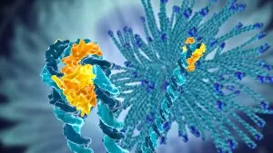
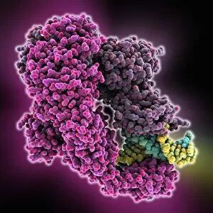
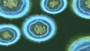
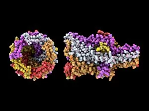
"Exploring the Intricate World Cells: A Fusion of Science and Art" Step into a mesmerizing realm where science meets art, as we delve into the captivating world cells. These tiny organisms, lacking a nucleus and other membrane-bound organelles, may seem simple at first glance, but their complexity is truly awe-inspiring. Let us begin our journey with Pyrococcus furiosus archaea artwork. This stunning portrayal captures the essence of these extremophiles that thrive in scorching temperatures. Their ability to survive in such harsh conditions showcases the resilience and adaptability of prokaryotes. Moving on to bacteria shapes depicted in F007 / 9891 artwork, we witness an array of diverse forms - from spirals to rods and spheres. Each shape represents a unique bacterial species, highlighting the remarkable diversity within this group. Zooming in further, we encounter molecular models showcasing the intricate structure of a 70S ribosome (F006 / 9651 & F006 / 9638). These essential cellular components play a crucial role in protein synthesis, underscoring their significance in prokaryotic biology. Next up is an artistic representation of a bacterial protease molecule (F006 / 9340). This enzyme's function lies in breaking down proteins within bacteria for various cellular processes. Its elegant structure reminds us that even at the microscopic level, beauty can be found. Shifting gears slightly, we explore DNA-bending protein molecular models that contribute to gene regulation and packaging (repeated twice). These proteins sculpt DNA strands into compact structures while ensuring proper gene expression – an exquisite dance between molecules. Lastly, Chlamydia infection artworks (C016 / 8952 & C016 / 8954) shed light on one aspect of prokaryotes' impact on human health. The illustrations depict how this sexually transmitted bacterium invades host cells, emphasizing the importance of understanding and combating such infections.