Fraud Blocker Skip to main content

Protein Shell Collection

Protein shells, the guardians of genetic material

Background imageProtein Shell Collection: Cucumber mosaic virus, computer model

Cucumber mosaic virus, computer model
Cucumber mosaic virus (CMV), computer model. This image was created using molecular modelling software and data from X-ray crystallography

Background imageProtein Shell Collection: Murine norovirus with antibody fragments

Murine norovirus with antibody fragments
Murine norovirus (MNV) with antibody fragments, computer model. This image was created using molecular modelling software and data from cryo- electron microscopy

Background imageProtein Shell Collection: HK97 bacteriophage capsid

HK97 bacteriophage capsid, molecular model. Bacteriophages are viruses that infect bacteria, in this case enterobacteria such as E. coli (Escherichia coli), with the phage head shown here

Background imageProtein Shell Collection: Chikungunya virus capsid

Chikungunya virus capsid, molecular model. This virus, transmitted by mosquitoes in tropical Africa and Asia, causes fever and joint pain in humans, similar to dengue fever

Background imageProtein Shell Collection: HK97 bacteriophage procapsid

HK97 bacteriophage procapsid. Molecular model showing the structure of the prohead-I procapsid of the HK97 bacteriophage. Bacteriophages are viruses that infect bacteria

Background imageProtein Shell Collection: Turnip yellow mosaic virus capsid

Turnip yellow mosaic virus capsid, molecular model. This virus infects a wide variety of plants, including crops such as turnips and cabbages, causing yellow patches on the leaves

Background imageProtein Shell Collection: Sindbis virus capsid, molecular model

Sindbis virus capsid, molecular model. This virus, transmitted by mosquitoes, causes sindbis fever in humans. In viruses, the capsid is the protein shell that encloses the genetic material

Background imageProtein Shell Collection: Murine polyomavirus capsid

Murine polyomavirus capsid, molecular model. This virus, one of a range named for their potential to cause multiple tumours, infects mice

Background imageProtein Shell Collection: Brome mosaic virus capsid

Brome mosaic virus capsid, molecular model. This plant virus infects grasses, especially brome grasses, and also barley. It causes mosaic patches of discolouration

Background imageProtein Shell Collection: Cowpea chlorotic mottle virus capsid

Cowpea chlorotic mottle virus capsid, molecular model. This virus (CCMV) infects the cowpea plant (Vigna unguiculata), causing yellow spots of discolouration

Background imageProtein Shell Collection: Avian polyomavirus capsid

Avian polyomavirus capsid, molecular model. This virus, one of a range named for their potential to cause multiple tumours, infects birds. Discovered in budgerigars in 1981, it is often fatal

Background imageProtein Shell Collection: Cytoplasmic polyhedrosis virus capsid

Cytoplasmic polyhedrosis virus capsid, molecular model. Part of the Cypovirus genus and invariably fatal, this insect virus is transmitted by contamination of leaves eaten (examples include silkworms)

Background imageProtein Shell Collection: Theilers encephalomyelitis virus capsid

Theilers encephalomyelitis virus capsid, molecular model. This virus, which causes brain and spinal cord inflammation in mice, is used in research

Background imageProtein Shell Collection: Tobacco necrosis virus capsid

Tobacco necrosis virus capsid, molecular model. This plant virus infects a wide rage of plants, including the tobacco plant for which it is named. The virus causes tissue death (necrosis)

Background imageProtein Shell Collection: Flock house virus capsid F006 / 9755

Flock house virus capsid F006 / 9755
Flock house virus capsid, molecular model. The flock house virus is a member of the Nodaviridae family. It kills the New Zealand grass grub insect

Background imageProtein Shell Collection: Bluetongue virus capsid, molecular model F006 / 9687

Bluetongue virus capsid, molecular model F006 / 9687
Bluetongue virus capsid, molecular model. This virus, transmitted by midges, causes bluetongue disease in ruminants (commonly sheep, and less frequently cattle, goats and deer)

Background imageProtein Shell Collection: Bacterial nanocompartment F006 / 9672

Bacterial nanocompartment F006 / 9672
Bacterial nanocompartment. Molecular molecule of a nanocompartment from the bacterium Thermotoga maritima. This is a shell formed from encapsulin proteins that encloses enzymes that defend the cell

Background imageProtein Shell Collection: Rous sarcoma virus capsid F006 / 9653

Rous sarcoma virus capsid F006 / 9653
Rous sarcoma virus capsid, molecular model. In viruses, the capsid is the protein shell that encloses the genetic material

Background imageProtein Shell Collection: Grapevine fanleaf virus capsid

Grapevine fanleaf virus capsid, molecular model. This plant virus is named for its infection of grape vines. It is transmitted by the nematode worm Xiphinema index

Background imageProtein Shell Collection: Mengovirus capsid, molecular model F006 / 9617

Mengovirus capsid, molecular model F006 / 9617
Mengovirus capsid, molecular model. A capsid consists of subunits called capsomeres that self-assemble to form the shell seen here

Background imageProtein Shell Collection: Hepatitis B virus capsid, molecular model F006 / 9594

Hepatitis B virus capsid, molecular model F006 / 9594
Hepatitis B virus capsid, molecular model. This virus, transmitted through infected bodily fluids or blood, causes the disease hepatitis B in humans, leading to acute liver inflammation

Background imageProtein Shell Collection: SV40 virus capsid, molecular model F006 / 9508

SV40 virus capsid, molecular model F006 / 9508
SV40 virus capsid, molecular model. Simian virus 40 (SV40) is found in monkeys such as Rhesus monkeys and macaques. Potentially tumour-causing, it is used in laboratory research and in vaccines

Background imageProtein Shell Collection: Parvovirus particle, molecular model F006 / 9499

Parvovirus particle, molecular model F006 / 9499
Parvovirus particle. Molecular model showing the structure of the capsid (outer protein coat) of a human parvovirus (family Parvoviridae) particle

Background imageProtein Shell Collection: Poliovirus type 3 capsid, molecular model F006 / 9465

Poliovirus type 3 capsid, molecular model F006 / 9465
Poliovirus type 3 capsid, molecular model. This enterovirus causes poliomyelitis (polio) in humans, which affects the nervous system, sometimes leading to paralysis

Background imageProtein Shell Collection: VEE equine encephalitis virus capsid

VEE equine encephalitis virus capsid
Venezuelan equine encephalitis virus capsid, molecular model. This mosquito-borne virus can kill horses and other equine species, causing brain and spinal cord inflammation

Background imageProtein Shell Collection: Semliki forest virus capsid F006 / 9297

Semliki forest virus capsid F006 / 9297
Semliki forest virus capsid, molecular model. This virus, named for the forest in Uganda where it was identified, is spread by the bite of mosquitoes. It can infect both humans and animals

Background imageProtein Shell Collection: Murine minute virus capsid

Murine minute virus capsid, molecular model. This parvovirus infects mice, its only known natural host. It is highly infectious, transmitted through the nose and mouth

Background imageProtein Shell Collection: SV40 virus capsid, molecular model C018 / 7904

SV40 virus capsid, molecular model C018 / 7904
SV40 virus capsid, molecular model. Simian virus 40 (SV40) is found in monkeys such as Rhesus monkeys and macaques. Potentially tumour-causing, it is used in laboratory research and in vaccines

Background imageProtein Shell Collection: SV40 virus capsid, molecular model C018 / 7903

SV40 virus capsid, molecular model C018 / 7903
SV40 virus capsid, molecular model. Simian virus 40 (SV40) is found in monkeys such as Rhesus monkeys and macaques. Potentially tumour-causing, it is used in laboratory research and in vaccines

Background imageProtein Shell Collection: Canine parvovirus capsid C015 / 8460

Canine parvovirus capsid C015 / 8460
Canine parvovirus capsid, molecular model. Canine parvovirus type 2 (CPV2) was first recognized in 1978 and spread worldwide in less than two years

Background imageProtein Shell Collection: Canine parvovirus capsid C015 / 8459

Canine parvovirus capsid C015 / 8459
Canine parvovirus capsid, molecular model. Canine parvovirus type 2 (CPV2) was first recognized in 1978 and spread worldwide in less than two years

Background imageProtein Shell Collection: Adeno-associated virus, molecular model C018 / 0449

Adeno-associated virus, molecular model C018 / 0449
Adeno-associated virus (aV), molecular model. This image shows the core protein of the non-enveloped virus that surrounds the genetic material

Background imageProtein Shell Collection: Hepatitis B virus, molecular model C018 / 0455

Hepatitis B virus, molecular model C018 / 0455
Hepatitis B virus core protein, molecular model. The virus causes hepatitis B, an inflammatory liver disease. The core proteins enclose the virus DNA and are in turn surrounded by a lipid envelope

Background imageProtein Shell Collection: Norovirus capsid, molecular model C018 / 0457

Norovirus capsid, molecular model C018 / 0457
Norovirus capsid, molecular model. Also known as the winter vomiting bug, Noroviruses cause gastroenteritis and are highly contagious, infecting approximately 267 million people a year

Background imageProtein Shell Collection: Astrovirus capsid, molecular model C018 / 0450

Astrovirus capsid, molecular model C018 / 0450
Astrovirus capsid, molecular model. This icosahedral virus was identified in 1975 using electron microscopy. It has a characteristic five-pointed symmetry to its surface, as seen here

Background imageProtein Shell Collection: Hepatitis E virus, molecular model C018 / 0445

Hepatitis E virus, molecular model C018 / 0445
Hepatitis E virus core protein, molecular model. The virus causes hepatitis E, an inflammatory liver disease that usually only lasts a few weeks

Background imageProtein Shell Collection: Human polio virus, molecular model

Human polio virus, molecular model
Human polio virus capsid, molecular model. Poliovirus causes poliomyelitis, a disease that can cause paralysis in up to 2 percent of patients, and in some cases death

Background imageProtein Shell Collection: Clathrin lattice, molecular model C018 / 0453

Clathrin lattice, molecular model C018 / 0453
Clathrin lattice, molecular model. This polyhedral protein lattice coats eukaryotic cell membranes (vesicles) and is involved in protein secretion and membrane trafficking

Background imageProtein Shell Collection: Clathrin lattice, molecular model C018 / 0452

Clathrin lattice, molecular model C018 / 0452
Clathrin lattice, molecular model. This polyhedral protein lattice coats eukaryotic cell membranes (vesicles) and is involved in protein secretion and membrane trafficking

Background imageProtein Shell Collection: Clathrin lattice, molecular model C018 / 0454

Clathrin lattice, molecular model C018 / 0454
Clathrin lattice, molecular model. This polyhedral protein lattice coats eukaryotic cell membranes (vesicles) and is involved in protein secretion and membrane trafficking

Background imageProtein Shell Collection: KSHV virus capsid, molecular model C018 / 0456

KSHV virus capsid, molecular model C018 / 0456
KSHV virus capsid, molecular model. KSHV is Kaposis sarcoma-associated herpesvirus. The virus is an oncovirus, which is a virus that can cause cancer

Background imageProtein Shell Collection: SV40 virus capsid, molecular model

SV40 virus capsid, molecular model. Simian virus 40 (SV40) is found in monkeys such as Rhesus monkeys and macaques. Potentially tumour-causing, it is used in laboratory research and in vaccines

Background imageProtein Shell Collection: Dengue virus capsid, molecular model

Dengue virus capsid, molecular model. This virus, transmitted by mosquito bites, causes the tropical disease dengue fever in humans

Background imageProtein Shell Collection: Ryegrass mottle virus capsid

Ryegrass mottle virus capsid, molecular model. This plant virus is named for its infection of ryegrass, and the discolouration it causes

Background imageProtein Shell Collection: Norwalk virus capsid, molecular model

Norwalk virus capsid, molecular model. This norovirus, which causes a viral form of gastroenteritis, is transmitted from person-to-person or through contaminated food

Background imageProtein Shell Collection: Semliki forest virus capsid

Semliki forest virus capsid, molecular model. This virus, named for the forest in Uganda where it was identified, is spread by the bite of mosquitoes. It can infect both humans and animals

Background imageProtein Shell Collection: Physalis mottle virus capsid

Physalis mottle virus capsid
Avian polyomavirus capsid, molecular model. This virus, one of a range named for their potential to cause multiple tumours, infects birds. Discovered in budgerigars in 1981, it is often fatal

Background imageProtein Shell Collection: Bombyx mori densovirus 1 capsid

Bombyx mori densovirus 1 capsid
Bombyx mori densovirus 1 (BmDNV-1), molecular model. This virus infects crustaceans and insects, especially the silkworm (Bombyx mori)


All Professionally Made to Order for Quick Shipping

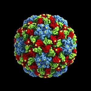
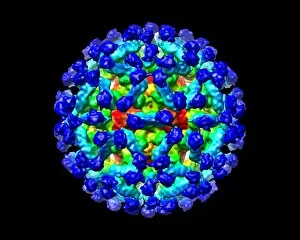
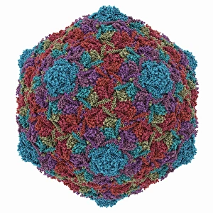
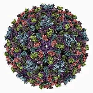
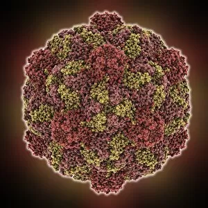
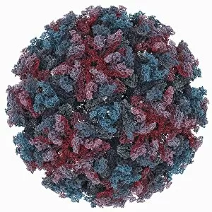
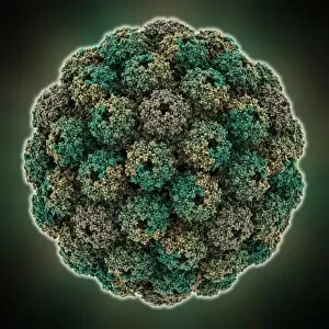
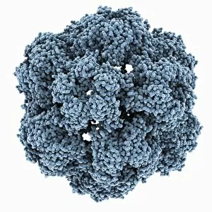
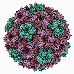
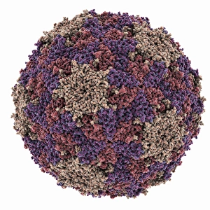
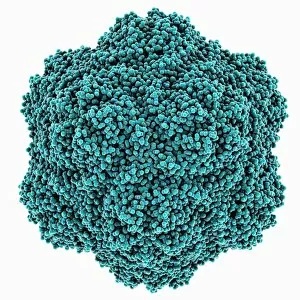
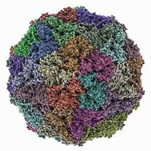
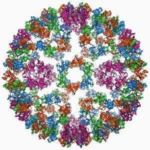
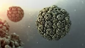
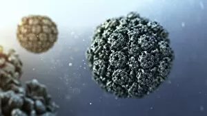
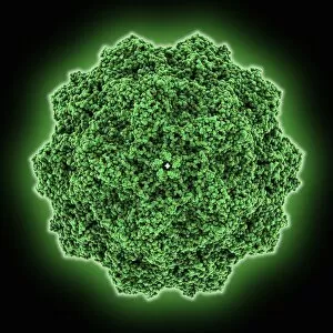
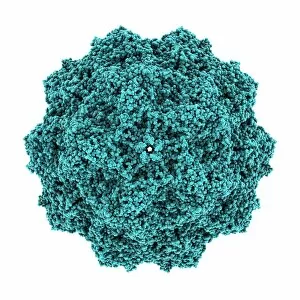
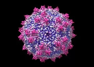
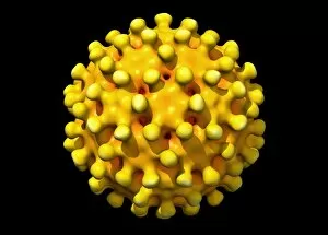
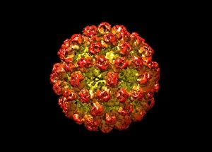
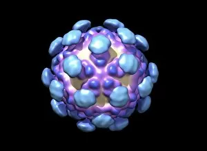
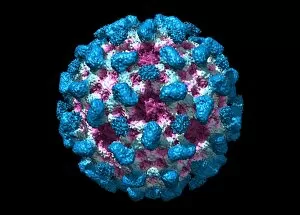
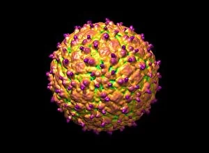
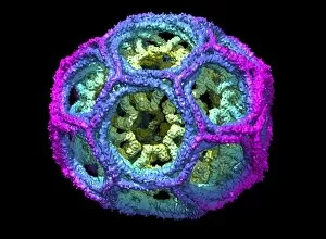
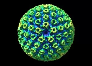
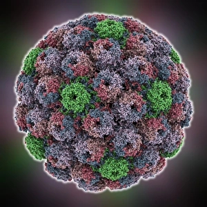
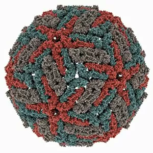
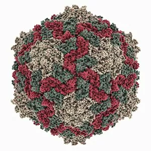
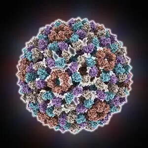
Protein shells, the guardians of genetic material. 🧬✨ From Bacteriophage phi29 to Simian immunodeficiency virus (SIV), these tiny marvels play a crucial role in protecting and delivering genetic information. Through computer models, scientists have unraveled their intricate structures and marveled at their functionality. Take a peek into the world of protein shells: Bacteriophage phi29: This remarkable phage boasts an elegant protein shell that encapsulates its DNA payload, ensuring safe delivery to host bacteria. Simian immunodeficiency virus (SIV): Its protein shell shields the viral RNA from external threats while facilitating its entry into host cells, leading to potential insights for HIV research. Murine norovirus with antibody fragments: The combination of this virus's capsid and antibody fragments holds promise for developing effective treatments against norovirus infections. HK97 bacteriophage capsid: With its unique shape and structure, this bacteriophage's protein shell acts as armor against host defenses during infection. Chikungunya virus capsid: A key player in chikungunya fever transmission, this virus's protein shell enables efficient replication within mosquito vectors and subsequent human infection. Turnip yellow mosaic virus capsid: Nature's architectural masterpiece. This plant-infecting virus showcases a beautifully symmetrical protein shell that safeguards its genetic material during transmission between plants. Hepatitis B virus capsid molecular model: Understanding the intricacies of this viral protein shell aids in designing antiviral strategies against hepatitis B infections worldwide. Infectious bursal disease virus capsid: A significant threat to poultry health, studying this avian pathogen's protective coat may pave the way for improved vaccines or treatment options. Sindbis virus capsid molecular model: By deciphering the structure of this alphavirus' outer layer, researchers gain insights into its replication process and potential targets for antiviral interventions.