Fraud Blocker Skip to main content

Proteomics Collection (page 38)

Proteomics, the study of proteins and their functions within an organism, is a fascinating field that unravels the intricate workings of life

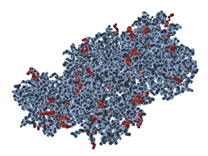
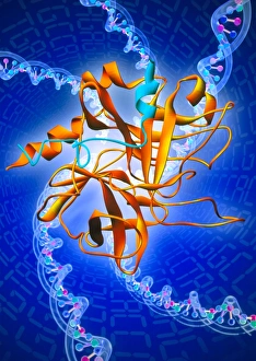
Background imageProteomics Collection: Trypsin molecule, computer artwork

Trypsin molecule, computer artwork
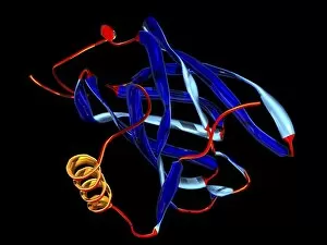
Trypsin molecule. Computer model of a molecule of the digestive enzyme trypsin. Trypsin is a complex protein, released by the pancreas to break down proteins into smaller chains of amino acids

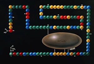
Background imageProteomics Collection: Art of structure of enzyme cytochrome C

Art of structure of enzyme cytochrome C
Cytochrome C enzyme. Illustration of a model of the enzyme cytochrome C, important in the process of respiration. Cytochromes are proteins often regarded as enzymes

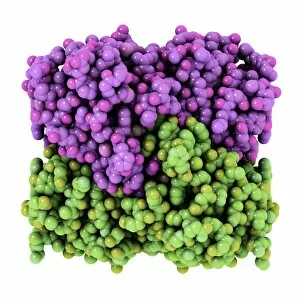
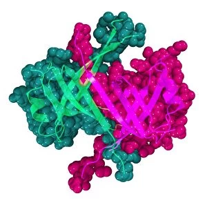
Background imageProteomics Collection: Cytidine deaminase, molecular model

Cytidine deaminase, molecular model
Cytidine deaminase. Computer model of the enzyme, activation-induced (cytidine) deaminase (AID). The tertiary structures of two protein complexes (purple and green)

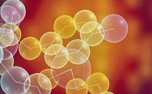
Background imageProteomics Collection: Encephalin peptide

Encephalin peptide
Encephalin. Computer molecular graphic of part of a molecule of encephalin, a polypeptide found in the human brain. It has a painkilling effect

Background imageProteomics Collection: Transcription factor-DNA, molecular model

Transcription factor-DNA, molecular model. Strand of DNA (deoxyribonucleic acid, yellow) being acted on by a transcription factor protein (red and green)

Background imageProteomics Collection: Preventing bacterial dormancy, artwork

Preventing bacterial dormancy, artwork. Proteins (red and yellow) being used to control bacterial DNA (blue, deoxyribonucleic acid) and prevent a mechanism known as bacterial dormancy

Background imageProteomics Collection: Influenza virus structure, artwork

Influenza virus structure, artwork
Influenza virus structure, cutaway artwork. The core of the virus is its genetic material, here 8 coloured ribbons of single-stranded RNA (ribonucleic acid)

Background imageProteomics Collection: Haemagglutinin flu proteins, artwork

Haemagglutinin flu proteins, artwork
Haemagglutinin flu proteins (red), artwork. Haemagglutinin is a membrane (surface) protein for the infuenza virus. An example is H1N1 flu virus, where H1 stands for haemagglutinin 1

Background imageProteomics Collection: Flu virion protein assembly, artwork

Flu virion protein assembly, artwork. Three types of flu membrane (surface) proteins are shown coming together here to form the coating for a new virion

Background imageProteomics Collection: Blocked flu virus ion channel, artwork

Blocked flu virus ion channel, artwork. This ion channel is an M2 membrane ion channel, found in the membranes of flu viruses

Background imageProteomics Collection: Pore forming bacterial toxin

Pore forming bacterial toxin. Computer model showing the molecular structure of the bacterial toxin pneumolysin. Pneumolysin is a pore forming toxin (PFT)

Background imageProteomics Collection: RNA processing protein, molecular model

RNA processing protein, molecular model
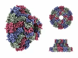
RNA processing protein, RNase MRP. Computer model showing the molecular structure of mitochondrial RNase MRP (mitochondrial RNA processing)

Background imageProteomics Collection: Molecules of haemocyanin protein

Molecules of haemocyanin protein
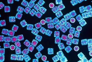
Haemocyanin. Coloured transmission electron micro- graph (TEM) of molecules of haemocyanin, a respiratory protein and pigment found in molluscs and arthropods

Background imageProteomics Collection: Microglobulin protein, molecular model

Microglobulin protein, molecular model
Microglobulin protein. Molecular models of two overlapping views of the protein beta-2 microglobulin. This protein, with a relatively small molecular mass

Background imageProteomics Collection: Molecular motor protein

Molecular motor protein. Computer model showing the structure of a two-headed motor protein, Myosin V. Motor proteins convert chemical energy into mechanical movements in response to specific

Background imageProteomics Collection: Mouse urinary protein, molecular model

Mouse urinary protein, molecular model
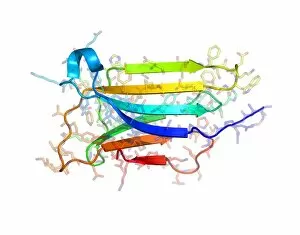
Mouse urinary protein (MUP), molecular model. MUP refers to a family of similar proteins found in mouse urine. The proteins act as pheromones, airborne chemical signals

Background imageProteomics Collection: Thrombin protein

Thrombin protein

Background imageProteomics Collection: Myoglobin protein

Myoglobin protein
Myoglobin. Computer model of the protein myoglobin that contains the iron-containing haem group (not seen). Myoglobin consists of a chain of 153 amino acids folded into a globin molecule that has a

Background imageProteomics Collection: RNA-editing enzyme combined with RNA

RNA-editing enzyme combined with RNA. Computer model showing the mRNA-editing enzyme, APOBEC-1 (apolipoprotein B mRNA editing enzyme, catalytic polypeptide 1)

Background imageProteomics Collection: RNA-binding protein, molecular model

RNA-binding protein, molecular model
RNA-binding protein. Computer model of the RNA-binding protein ACF (APOBEC-1 complementation factor). It is thought that ACF functions as an RNA-binding subunit that docks APOBEC-1 with an RNA

Background imageProteomics Collection: Protozoan RNA-binding protein complex

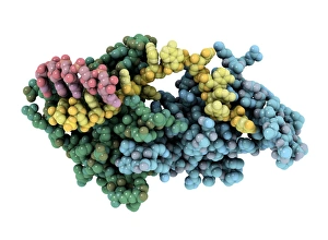
Protozoan RNA-binding protein complex
RNA-binding protein complex. Computer model showing a guide RNA-binding protein complex (green and blue), bound to guide RNA (gRNA, yellow and red))

Background imageProteomics Collection: Collagen fibre, molecular model

Collagen fibre, molecular model. Collagen is a long structural protein, which usually takes the form of a triple helix known as tropocollagen

Background imageProteomics Collection: Pepsin molecule

Pepsin molecule
Pepsin enzyme. Computer graphic of the protein- digesting enzyme pepsin. It is a protease enzyme that is secreted as part of gastric juice into the stomach in an inactive form known as pepsinogen

Background imageProteomics Collection: Fibroblast growth factor receptor 2

Fibroblast growth factor receptor 2 (FGFR2). Molecular models of the secondary structure (top) and the tertiary structure (bottom) of FGFR2

Background imageProteomics Collection: Single stranded DNA-binding protein

Single stranded DNA-binding protein (SSBP). Molecular model showing the secondary and tertiary structures of a protein that binds to the single stranded DNA (deoxyribonucleic acid)

Background imageProteomics Collection: Enzyme catalysing DNA recombination

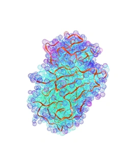
Enzyme catalysing DNA recombination. Computer model of the enzyme flippase recombinase (FLP recombinase, atoms represented as tubes)

Background imageProteomics Collection: Bacteriophage DNA packaging motor

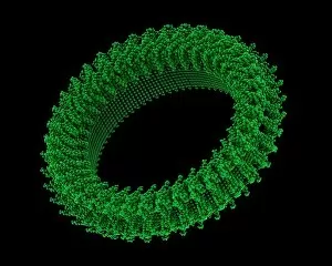
Bacteriophage DNA packaging motor, molecular model. Bacteriophages are viruses that only infect bacteria. They enter the host cell (a bacterium)

Background imageProteomics Collection: Nerve growth factor, molecular model

Nerve growth factor, molecular model
Nerve growth factor. Molecular model showing the secondary structure of nerve growth factor (NGF). NGF is a small protein, which is involved in the growth

Background imageProteomics Collection: Nitrogenase protein, molecular model

Nitrogenase protein, molecular model
Nitrogenase protein. Molecular model of the MoFe protein, one of two proteins (MoFe and Fe) that combine to form the enzyme nitrogenase

Background imageProteomics Collection: Advanced Light Source synchrotron

Advanced Light Source synchrotron
Advanced Light Source (ALS) synchrotron for producing intense X-rays used to determine the structures of proteins at Lawrence Berkeley Laboratory, USA


All Professionally Made to Order for Quick Shipping

Proteomics, the study of proteins and their functions within an organism, is a fascinating field that unravels the intricate workings of life. From anaesthetics inhibiting ion channels to immunoglobulin G antibody molecules, proteomics delves into the molecular mechanisms that shape our existence. In the realm of brain research, scientists explore how proteins influence cognition and behavior. They investigate DNA nucleosomes' structure and function, unraveling their role in gene regulation. Antibodies take center stage as artwork showcases their diverse forms and crucial role in immune defense. Zinc fingers bound to a DNA strand highlight protein-DNA interactions critical for genetic processes. Meanwhile, manganese superoxide dismutase enzyme aids in protecting cells from oxidative stress. The SARS coronavirus protein becomes a subject of intense scrutiny as researchers strive to understand its pathogenicity. Cytochrome b5 molecule reveals insights into electron transfer reactions within cells while glutamine synthetase enzyme plays a vital role in nitrogen metabolism. Lastly, RNA-editing enzymes offer potential therapeutic targets for various diseases with their ability to modify genetic information at the RNA level. Through proteomics, we unlock nature's secrets one protein at a time - deciphering their structures, unraveling their functions, and ultimately enhancing our understanding of life itself.