Fraud Blocker Skip to main content

Protozoa Collection (page 3)

Protozoa, the microscopic wonders of nature

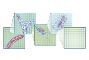
Background imageProtozoa Collection: Cross section biomedical illustration on grid of Worm, Fungi, Protozoa

Cross section biomedical illustration on grid of Worm, Fungi, Protozoa, and Bacteria infection and infestation

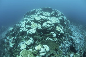
Background imageProtozoa Collection: Corals are beginning to bleach on a reef in Indonesia

Corals are beginning to bleach on a reef in Indonesia. Bleaching occurs as corals expel their endosymbionts (zooxanthellae) living within their tissues

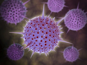
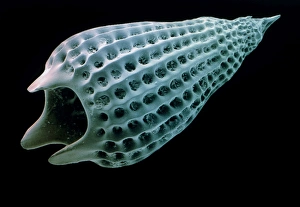
Background imageProtozoa Collection: Conceptual image of Radiolarians with a skeletal frame

Conceptual image of Radiolarians with a skeletal frame. Radiolarians are tiny protozoans that live in the ocean

Background imageProtozoa Collection: Conceptual image of paramecium

Conceptual image of paramecium

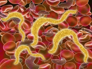
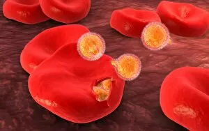
Background imageProtozoa Collection: African trypanosomiasis in the red blood cells

African trypanosomiasis in the red blood cells. African trypanosomiasis, also known as sleeping sickness, is a parasitic infection caused by protozoa

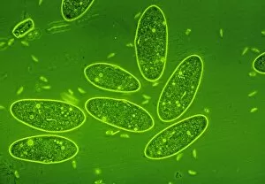
Background imageProtozoa Collection: Microscopic view of paramecium

Microscopic view of paramecium

Background imageProtozoa Collection: Conceptual image of plasmodium causing malaria

Conceptual image of plasmodium causing malaria

Background imageProtozoa Collection: Conceptual image of malaria parasites within red blood cells

Conceptual image of malaria parasites within red blood cells

Background imageProtozoa Collection: Conceptual image of plasmodium

Conceptual image of plasmodium

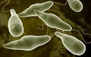
Background imageProtozoa Collection: Conceptual image of Euglena

Conceptual image of Euglena

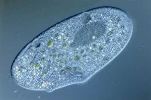
Background imageProtozoa Collection: Microscopic view of Paramecium bursaria

Microscopic view of Paramecium bursaria, a species of ciliate protozoan that has a mutualistic symbiotic relationship with green algae called Zoochlorella

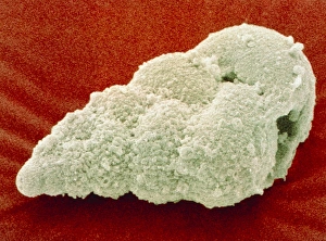
Background imageProtozoa Collection: Microscopic view of protozoa

Microscopic view of protozoa. Leishmaniasis and dysentery are examples of diseases involving protozoa, often transmitted to humans by the bite of a parasite-carrying insect

Background imageProtozoa Collection: Conceptual image of Trypanosoma

Conceptual image of Trypanosoma

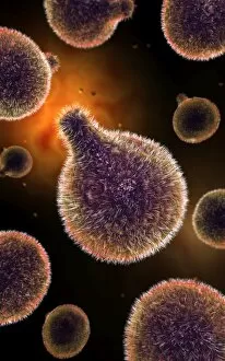
Background imageProtozoa Collection: Microscopic view of a microbe

Microscopic view of a microbe. Microbes are single-cell organisms so tiny that millions can fit into the eye of a needle

Background imageProtozoa Collection: Malria spores in the human blood stream

Malria spores in the human blood stream
The malaria spores move from the mosquito to the human blood stream. the spores divide many times and produce large numbers of spores

Background imageProtozoa Collection: Typical enlarged spleen of a Malaria patient. Malaria is caused by a parasitic protozoa

Typical enlarged spleen of a Malaria patient. Malaria is caused by a parasitic protozoa transmitted by the Anopheles mosquito, Until the 1930s the only effective treatment was the drug Quinine

Background imageProtozoa Collection: Head of the Anopheles Mosquito showing thefemale mouthparts. The bite of the female

Head of the Anopheles Mosquito showing thefemale mouthparts. The bite of the female of this species can transmit the Malaria parasite. Engraving, 1912

Background imageProtozoa Collection: Crowds on the streets in Germany during the political risings of 1918-1920

Crowds on the streets in Germany during the political risings of 1918-1920

Background imageProtozoa Collection: Model of vorticella

Model of vorticella

Background imageProtozoa Collection: Large group of Flagellates, illustration

Large group of Flagellates, illustration
Biology - Protozoa - Large group of Flagellates, illustration

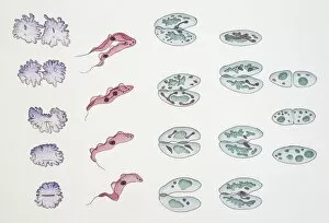
Background imageProtozoa Collection: Division of Protozoa cells, illustration

Division of Protozoa cells, illustration Biology

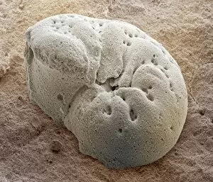
Background imageProtozoa Collection: Foraminiferan fossil, SEM

Foraminiferan fossil, SEM
Foraminiferan fossil. Coloured scanning electron micrograph (SEM) of the fossilised shell (test) of a foraminiferan. Foraminifera are single-celled marine protozoa that construct

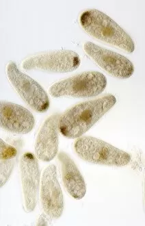
Background imageProtozoa Collection: LM of the protozoan Paramecium sp

LM of the protozoan Paramecium sp
Light micrograph of the ciliate protozoan, Paramecium sp. They are one of the most highly- evolved of all unicellular animals, and are plentiful in fresh water ponds

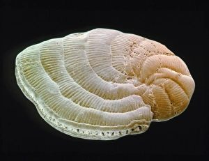
Background imageProtozoa Collection: Coloured SEM of foraminiferan, Peneroplis sp

Coloured SEM of foraminiferan, Peneroplis sp
Foraminiferan shell. Coloured scanning electron micrograph (SEM) of the test (shell) of the Mediterranean foraminiferan, Peneroplis proteus

Background imageProtozoa Collection: SEM of Paramecium

SEM of Paramecium
Paramecium. Coloured scanning electron micrograph (SEM) of the single-celled protozoan, Paramecium caudatum. This ciliate animal has cilia on its cell membrane surface enabling it to swim with an

Background imageProtozoa Collection: LM of of a single Paramecium sp

LM of of a single Paramecium sp
Light micrograph of the ciliate protozoan Paramecium sp. The internal structure of the organism is particularly clear. The large dark area toward the top of the frame is the meganucleus

Background imageProtozoa Collection: Coloured SEM of Podocyrtis mitra, a radiolarian

Coloured SEM of Podocyrtis mitra, a radiolarian
Radiolarian. Coloured scanning electron micrograph (SEM) of the test (skeleton) of a Podocyrtis mitra, a type of radiolarian protozoan

Background imageProtozoa Collection: LM of paramecium sp

LM of paramecium sp
Light micrograph in phase contrast illumination of the ciliate protozoan, Paramecium sp. The internal structure of Paramecium is visible here with many food and water vacuoles seen as bright spots

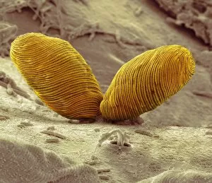
Background imageProtozoa Collection: Protozoan sexual reproduction, SEM Z105 / 0240

Protozoan sexual reproduction, SEM Z105 / 0240
Protozoan sexual reproduction. Coloured scanning electron micrograph (SEM) of two Paramecium caudatum protozoa reproducing sexually by a method called conjugation

Background imageProtozoa Collection: Coloured SEM of foraminiferan, Elphidium sp

Coloured SEM of foraminiferan, Elphidium sp
Foraminiferan shell. Coloured scanning electron micrograph (SEM) of the test (shell) of the British formaminiferan, Elphidium crispum

Background imageProtozoa Collection: Coloured SEM of Foraminifera Textularia sp. fossil

Coloured SEM of Foraminifera Textularia sp. fossil
Fossil foraminifera. Coloured scanning electron micrograph (SEM) of the fossil shell of the single-celled protozoan Textularia sp. belonging to the order Foraminifera

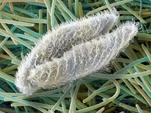
Background imageProtozoa Collection: Paramecium protozoa, SEM Z105 / 0239

Paramecium protozoa, SEM Z105 / 0239
Paramecium protozoa. Coloured scanning electron micrograph (SEM) of two Paramecium caudatum protozoa on filaments (green) of Oscillatoria sp. blue-green algae. P

Background imageProtozoa Collection: Calcareous phytoplankton fossil, SEM Z100 / 0212

Calcareous phytoplankton fossil, SEM Z100 / 0212
Calcareous phytoplankton fossil. Coloured scanning electron micrograph (SEM) of a fossil of the skeleton (coccosphere) of a coccolithotrope, a small marine algal organism

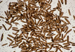
Background imageProtozoa Collection: Chilodonella ciliate protozoa, LM

Chilodonella ciliate protozoa, LM
Chilodonella protozoa. Light micrograph of several Chilodonella sp. ciliate protozoa. Chilodonella is a single-celled animal that is found in freshwater

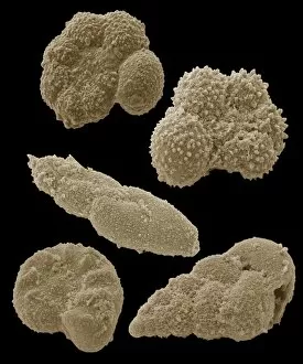
Background imageProtozoa Collection: Fossilised foraminiferans, SEM

Fossilised foraminiferans, SEM
Fossilised foraminiferans. Coloured scanning electron micrograph (SEM) of the fossilised shells of assorted foraminiferan protozoa (order Foraminifera)

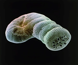
Background imageProtozoa Collection: Coloured SEM of foraminiferan, Spirolina sp

Coloured SEM of foraminiferan, Spirolina sp
Foraminiferan shell. Coloured scanning electron micrograph (SEM) of the test (shell) of the Mediterranean foraminiferan, Spirolina areitinus

Background imageProtozoa Collection: Foraminiferan tests

Foraminiferan tests (shells), macrophotograph. Species seen here include Bulimina gibba, Elphidium crispum, E. articulatum, Lagena sulcata, and Miliolinella circularis

Background imageProtozoa Collection: Foraminiferan fossil, SEM Z110 / 0239

Foraminiferan fossil, SEM Z110 / 0239
Foraminiferan fossil. Coloured scanning electron micrograph (SEM) of the fossilised shell (test) of a foraminiferan. Foraminifera are single-celled marine protozoa that construct

Background imageProtozoa Collection: Holosticha ciliate protozoan, SEM

Holosticha ciliate protozoan, SEM
Holosticha ciliate protozoan. Coloured scanning electron micrograph (SEM) of a Holosticha sp. ciliate protozoan ingesting diatoms (blue). Holosticha is a tiny single-celled aquatic organism

Background imageProtozoa Collection: Euplotes protozoa, SEM C016 / 9040

Euplotes protozoa, SEM C016 / 9040
Euplotes protozoa. Coloured scanning electron micrograph (SEM) showing the upperside (bottom) and underside (top) of two Euplotes sp. hypotrich ciliate protozoa

Background imageProtozoa Collection: Climacostomum protozoan, SEM C016 / 9063

Climacostomum protozoan, SEM C016 / 9063
Climacostomum protozoan. Coloured scanning electron micrograph (SEM) of a Climacostomum sp. unicellular ciliate protozoan, showing the cells large oral apparatus (round, centre)

Background imageProtozoa Collection: Euglena flagellate protozoa, SEM C016 / 9103

Euglena flagellate protozoa, SEM C016 / 9103
Euglena flagellate protozoa. Coloured scanning electron micrograph (SEM) of two Euglena sp. flagellate protozoa. Euglena sp

Background imageProtozoa Collection: Climacostomum protozoan, SEM C016 / 9121

Climacostomum protozoan, SEM C016 / 9121
Climacostomum protozoan. Coloured scanning electron micrograph (SEM) of a Climacostomum sp. unicellular ciliate protozoan, showing the cells large oral apparatus (round, centre)

Background imageProtozoa Collection: Climacostomum protozoan, SEM C016 / 9061

Climacostomum protozoan, SEM C016 / 9061
Climacostomum protozoan. Coloured scanning electron micrograph (SEM) of a Climacostomum sp. unicellular ciliate protozoan, showing the cells large oral apparatus (round, upper right)

Background imageProtozoa Collection: Climacostomum protozoan, SEM C016 / 9120

Climacostomum protozoan, SEM C016 / 9120
Climacostomum protozoan. Coloured scanning electron micrograph (SEM) of a Climacostomum sp. unicellular ciliate protozoan, showing the cells large oral apparatus (round, centre)

Background imageProtozoa Collection: Climacostomum protozoan, SEM C016 / 9065

Climacostomum protozoan, SEM C016 / 9065
Climacostomum protozoan. Coloured scanning electron micrograph (SEM) of a Climacostomum sp. unicellular ciliate protozoan, showing the cells large oral apparatus (round, upper right)

Background imageProtozoa Collection: Euglena flagellate protozoa, SEM C016 / 9104

Euglena flagellate protozoa, SEM C016 / 9104
Euglena flagellate protozoa. Coloured scanning electron micrograph (SEM) of two Euglena sp. flagellate protozoa. Euglena sp

Background imageProtozoa Collection: Climacostomum protozoan, SEM C016 / 9064

Climacostomum protozoan, SEM C016 / 9064
Climacostomum protozoan. Coloured scanning electron micrograph (SEM) of a Climacostomum sp. unicellular ciliate protozoan, showing the cells large oral apparatus (round, centre)


All Professionally Made to Order for Quick Shipping

Protozoa, the microscopic wonders of nature. From calcareous phytoplankton fossils to intricate artwork depicting Trypanosome protozoans, these single-celled organisms never cease to amaze. Take a closer look through the lens of SEM Z100 / 0213 and discover the mesmerizing beauty of their world. In one frame, we witness Plasmodium sp. , a malarial parasite that wreaks havoc on human health. Its complex structure reminds us of the delicate balance between life and disease. But not all it can harmful; many lead fascinating lives as scavengers or nutrient absorbers. Through light micrograph LM, we observe a kidney-shaped ciliate surrounded by Euglena sp. , showcasing their feeding habits at an astonishing magnification of x900. Diatoms, another group of protozoa, steal our attention with their intricate patterns and shapes. Acrosphaera radiolarian captivates us in its SEM image, displaying its ornate exoskeleton that protects it from predators. Foraminiferan tests (shells) take center stage in another SEM capture. These tiny structures tell stories about ancient oceans and provide valuable insights into Earth's history. Moving away from imagery, we encounter models representing foraminifera - miniature replicas that aid scientists in understanding these remarkable creatures' behavior and evolution. Not limited to aquatic environments alone, some protozoa find residence within our own bodies as intestinal parasites. TEM images reveal their presence while reminding us to prioritize hygiene and health practices. Amongst them is Trichomonas vaginalis captured through TEM - a reminder that even seemingly harmless organisms can cause discomfort if left unchecked. Lastly, Cryptosporidium protozoa appear under TEM's watchful eye – highlighting the importance of water safety measures due to this organism's ability to contaminate drinking sources with potential health risks. Protozoa may be small in size, but their impact on our world is immense.