
Pulvillus Collection
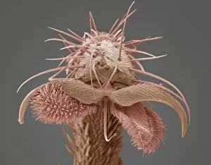
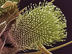
The intricate world of insect feet is revealed through scanning electron microscopy (SEM), showcasing the fascinating structures known as pulvilli


All Professionally Made to Order for Quick Shipping
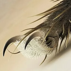
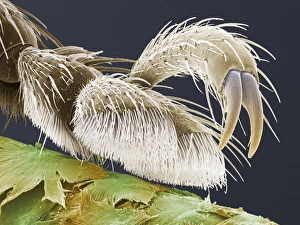
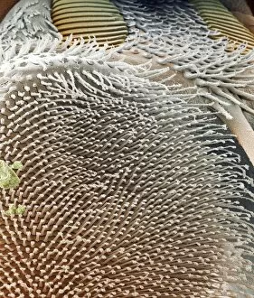
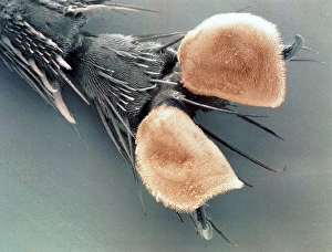
The intricate world of insect feet is revealed through scanning electron microscopy (SEM), showcasing the fascinating structures known as pulvilli. These tiny appendages, found on various fly species such as houseflies, vine weevils, fruit flies, cluster flies, grey flesh flies, yellow dung flies, gnats and more, play a crucial role in their daily lives. Examining the foot of a housefly under SEM reveals the delicate pulvillus that aids in its ability to cling onto surfaces effortlessly. Similarly, the SEM image of a vine weevil's foot showcases its unique pulvillus structure designed for gripping leaves and stems with precision. Moving on to fruit flies and cluster flies; their feet exhibit remarkable patterns when magnified by SEM. The ventral foot pads and claws of a grey flesh fly are particularly intriguing - these specialized structures allow them to walk upside down or even on smooth surfaces without slipping. Yellow dung flies also possess impressive pulvilli that assist them in navigating challenging terrains while searching for food sources. The detailed SEM images highlight how these insects adapt to their environments with astonishing precision. Further exploration into different fly species reveals repeated instances of pulvilli being captured under SEM - an indication of just how vital this feature is across various types of insects. Whether it be the minute details seen on gnat feet or the distinctive patterns observed within each specimen's pulvillus structure; every image provides valuable insights into nature's incredible adaptations. Studying these captivating images obtained through scanning electron microscopy allows us to appreciate the complexity and diversity present within insect feet. Pulvilli serve as essential tools enabling insects to thrive in their respective habitats – an extraordinary testament to evolution's ingenuity at work.