Fraud Blocker Skip to main content

Radiology Collection

Radiology, the art of peering into the human body's intricate mysteries, reveals a world unseen

Background imageRadiology Collection: Headache, X-ray artwork

Headache, X-ray artwork
Headache. Computer X-ray artwork of a lateral view of a human skull. The red area represents pain caused by a headache (cephalalgia)

Background imageRadiology Collection: Panoramic dental X-ray

Panoramic dental X-ray showing all the teeth in the upper and lower jaws

Background imageRadiology Collection: Assortment of coloured X-rays and body scans

Assortment of coloured X-rays and body scans
X-ray diagnosis. Assortment of coloured X-rays, body and brain scans. Parts of the body X-rayed include the skull and pelvis (at top), shoulder and lungs (upper centre and right)

Background imageRadiology Collection: Hands, X-ray

Hands, X-ray

Background imageRadiology Collection: Diagnostic Radiology, X-ray of two frogs, 1896

Diagnostic Radiology, X-ray of two frogs, 1896
UIG5086339 Diagnostic Radiology, X-ray of two frogs, 1896 by Eder, Josef Maria (1855 - 1944); (add.info.: Diagnostic Radiology, X-rays X-ray of two frogs, 1896)

Background imageRadiology Collection: Healthy knee, X-ray

Healthy knee, X-ray
Healthy knee. Coloured frontal (right) and profile (left) X-rays of the healthy knee of a 24 year old patient

Background imageRadiology Collection: Bacterial meningitis, MRI scan

Bacterial meningitis, MRI scan
Bacterial meningitis. Coloured magnetic resonance imaging (MRI) scan of an axial section through the brain of a 16-year-old patient

Background imageRadiology Collection: Tension pneumothorax, X-ray

Tension pneumothorax, X-ray
Tension pneumothorax. X-ray of the chest of a patient with tension pneumothorax that has lead to soft-tissue emphysema. Tension pneumothorax is a progressive deterioration

Background imageRadiology Collection: Picture No. 11676149

Picture No. 11676149
Girl Head x-ray and silhouette double exposure Date:

Background imageRadiology Collection: Total knee replacement, X-rays

Total knee replacement, X-rays
Total knee replacement. Coloured frontal (left) and profile (right) X-rays of the right knee of a 69 year old patient after total knee replacement surgery

Background imageRadiology Collection: French Auto Laboratory of Radiology, 31 Oct 1914. Creator: Bain News Service

French Auto Laboratory of Radiology, 31 Oct 1914. Creator: Bain News Service
French Auto Laboratory of Radiology, 31 Oct 1914 (date created or published later). A mobile radiology automobile during World War I, probably in France.

Background imageRadiology Collection: Healthy ankle joint, X-ray

Healthy ankle joint, X-ray
Healthy ankle joint. Coloured profile X-ray of the left ankle of a 21 year old patient. Strapping around the ankle is visible on this X-ray

Background imageRadiology Collection: Scoliosis of the spine, X-ray

Scoliosis of the spine, X-ray
Scoliosis of the spine. Coloured X-ray of the chest of a 31 year old patient with scoliosis (sideways curvature) of the spine

Background imageRadiology Collection: A radiologist takes a radiograph of a patient with the Microscope 500

A radiologist takes a radiograph of a patient with the Microscope 500. Instrument, discovered in America, which is much more protective to radiation. America. Date of Photograph:1950 - 1960

Background imageRadiology Collection: X-ray - Soldier with Bullet Bound Being Examined 1900

X-ray - Soldier with Bullet Bound Being Examined 1900
Wounded soldier from the Boer War been examined in a London hospital by means of X-ray. One of the interesting features of the Boer War was the demonstration of the practical utility of the X-rays

Background imageRadiology Collection: Wilhelm Konrad Roentgen (1845-1923) c. 1896 (b / w photo)

Wilhelm Konrad Roentgen (1845-1923) c. 1896 (b / w photo)
CHT237289 Wilhelm Konrad Roentgen (1845-1923) c.1896 (b/w photo) by German Photographer, (19th century); Musee du Centre Antoine Beclere, Paris

Background imageRadiology Collection: Wilhelm Konrad Roentgen (1845-1923) x-raying a young boy, from a book by Hans Kraemer, c

Wilhelm Konrad Roentgen (1845-1923) x-raying a young boy, from a book by Hans Kraemer, c
CHT237292 Wilhelm Konrad Roentgen (1845-1923) x-raying a young boy, from a book by Hans Kraemer, c.1898-99 (b/w photo) by German School, (19th century); Bibliotheque Nationale, Paris

Background imageRadiology Collection: Beach idyll in Rontgen style, cartoon about X-rays, c. 1896-99 (colour engraving)

Beach idyll in Rontgen style, cartoon about X-rays, c. 1896-99 (colour engraving)
CHT164182 Beach idyll in Rontgen style, cartoon about X-rays, c.1896-99 (colour engraving) by German School, (19th century); Bibliotheque des Arts Decoratifs, Paris

Background imageRadiology Collection: Radiguet x-ray machine (engraving) (b / w photo)

Radiguet x-ray machine (engraving) (b / w photo)
CHT237293 Radiguet x-ray machine (engraving) (b/w photo) by French School, (19th century); Bibliotheque Nationale, Paris, France; eArchives Charmet; French, out of copyright

Background imageRadiology Collection: Inside an early x-ray room (b / w photo)

Inside an early x-ray room (b / w photo)
STC343848 Inside an early x-ray room (b/w photo) by German Photographer, (19th century); Private Collection; (add.info.: X-rays were discovered by Wilhelm Conrad RAontgen (1845-1923) in 1895

Background imageRadiology Collection: Normal legs, X-rays

Normal legs, X-rays
Normal legs. Coloured frontal X-rays of the healthy legs, of a 35-year-old

Background imageRadiology Collection: Salivary gland stone, X-ray

Salivary gland stone, X-ray
Salivary gland stone. X-ray of the jaw of a 36-year-old patient with a stone (white, lower left) in the right submaxillary gland

Background imageRadiology Collection: Ischaemia, digital angiogram

Ischaemia, digital angiogram
image d angiographie numerisee( non soustraite) de la main gauche, en vue de face, par catheterisme hyperselectif des arteres du membre superieur gauche, sous anasthesie generale

Background imageRadiology Collection: Arthrosis of the hand, X-ray F006 / 4616

Arthrosis of the hand, X-ray F006 / 4616
Coloured X-ray of the hand of a patient with arthrosis, particularly the saddle joint. Arthrosis is a degenerative joint disease that damages cartilage and bone

Background imageRadiology Collection: Healthy knee, X-ray F006 / 9125

Healthy knee, X-ray F006 / 9125
Healthy knee. Coloured frontal (right) and profile (left) X-rays of the healthy knee of a 24 year old patient

Background imageRadiology Collection: Brain aneurysm, 3D scan

Brain aneurysm, 3D scan
Brain aneurysm. 3D computed tomography (CT) angiogram coupled with a magnetic resonance imaging (MRI) scan of the brain of a 38-year-old, showing a large aneurysm (bright)

Background imageRadiology Collection: Tuberculosis of the spine, MRI scan

Tuberculosis of the spine, MRI scan
Tuberculosis of the spine. Magnetic resonance imaging (MRI) scan of a sagittal section through the spine of a 74-year-old patient with tuberculosis (TB) of the spine, or Potts disease

Background imageRadiology Collection: Spondylolisthesis, 3D CT scan

Spondylolisthesis, 3D CT scan
Spondylolisthesis. Coloured 3D computed tomography (CT) scan of a section through the lumbar (lower back) spine of a 39-year-old patient with spondylolisthesis

Background imageRadiology Collection: Vascular system of the head, MRI scan

Vascular system of the head, MRI scan
Vascular system of the head. Magnetic resonance imaging (MRI) angiogram of a sagittal section through the head of a healthy 43-year-old, showing the blood vessels (white) of the brain and neck

Background imageRadiology Collection: Cardiac lymphoma, MRI scans

Cardiac lymphoma, MRI scans
Brain tumour. Coloured magnetic resonance imaging (MRI) scans of axial sections through the chest of a 22-year-old patient, showing a tumorous lesion invading the right ventricle of the heart (large)

Background imageRadiology Collection: Throat cancer, X-rays

Throat cancer, X-rays
Throat cancer. Coloured X-rays showing the transit of a contrast medium (dense white) through the throat of a 54-year-old patient with an adenocarcinoma (cancer) of the lower oesophagus (gullet)

Background imageRadiology Collection: Human head, X-ray artwork

Human head, X-ray artwork
Human head. Computer artwork of an X-ray through a human head, showing the brain (grey, top), cerebellum (blue), and brainstem and spinal cord (green)

Background imageRadiology Collection: Balloon angioplasty, X-ray

Balloon angioplasty, X-ray
Balloon angioplasty. Coloured X-ray of the abdomen of a 45-year-old patient with a history of thrombosis (blood clots) in the inferior vena cava (vein), which have narrowed (stenosed)

Background imageRadiology Collection: Deep brain stimulation, X-ray

Deep brain stimulation, X-ray
Image Xrays Crane (Sur Projection 3D Modèle) En Incidence De Profil Chez Une Patiente De 54 Ans, Parkinsonienne Traitee Sous Protocole De Neurochirurgie; Visualisation Des Electrodes De

Background imageRadiology Collection: Lung abscess, X-ray

Lung abscess, X-ray
Lung abscess. Coloured X-ray showing an abscess (dense coloured area, upper right) in the upper lobe of a patients left lung (right). An abscess is an accumulation of puss within a cavity in tissue

Background imageRadiology Collection: Full bladder, X-ray cystography

Full bladder, X-ray cystography
Full bladder. X-ray cystogram of the pelvic region of a 50-year-old incontinent patient, showing urine (white) leaking (lower centre) from the bladder (round, centre) during a stress test

Background imageRadiology Collection: Black and white illustration of a patient undergoing radiation therapy

Black and white illustration of a patient undergoing radiation therapy

Background imageRadiology Collection: X-ray image of a human jaw with treated teeth, Germany, Europe

X-ray image of a human jaw with treated teeth, Germany, Europe

Background imageRadiology Collection: Cancer treatment, artwork F007 / 4199

Cancer treatment, artwork F007 / 4199
Cancer treatment, conceptual image. Computer artwork representing the use of radiation in tumour treatment. Tumours are caused by the uncontrolled growth of previously normal cells

Background imageRadiology Collection: Arthrosis of the hand, X-ray F006 / 4605

Arthrosis of the hand, X-ray F006 / 4605
Coloured X-ray of the hand of a patient with arthrosis, particularly the saddle joint. Arthrosis is a degenerative joint disease that damages cartilage and bone

Background imageRadiology Collection: Arthrosis of the hand, X-ray F006 / 4595

Arthrosis of the hand, X-ray F006 / 4595
Coloured X-ray of the hand of a patient with arthrosis of the hands. particularly the saddle joint. Arthrosis is a degenerative joint disease that damages cartilage and bone

Background imageRadiology Collection: Arthrosis of the hand, X-ray F006 / 4598

Arthrosis of the hand, X-ray F006 / 4598
Coloured X-ray of the hand of a patient with arthrosis, particularly the saddle joint. Arthrosis is a degenerative joint disease that damages cartilage and bone

Background imageRadiology Collection: Hands, X-ray F006 / 8801

Hands, X-ray F006 / 8801
Hands, X-ray

Background imageRadiology Collection: Coronary stent, X-ray F006 / 9141

Coronary stent, X-ray F006 / 9141
Coronary stent. Coloured angiogram (blood vessel X-ray) of the coronary arteries of a 52 year old patient. The artery at right has had a stent placed in it to treat a blockage

Background imageRadiology Collection: Normal mammogram F006 / 9136

Normal mammogram F006 / 9136
Normal mammogram. Coloured X-ray of the left breast (mammogram) of a 27 year old patient

Background imageRadiology Collection: Knee disease, X-ray F006 / 9138

Knee disease, X-ray F006 / 9138
Knee disease. Coloured X-ray of the right knee of an 18 year old patient with osteochondritis dissecans (OCD) of the knee

Background imageRadiology Collection: Normal mammogram F006 / 9135

Normal mammogram F006 / 9135
Normal mammogram. Coloured X-ray of the left breast (mammogram) of a 27 year old patient

Background imageRadiology Collection: Scoliosis of the spine, X-ray F006 / 9134

Scoliosis of the spine, X-ray F006 / 9134
Scoliosis of the spine. Coloured X-ray of the chest of a 31 year old patient with scoliosis (sideways curvature) of the spine


All Professionally Made to Order for Quick Shipping

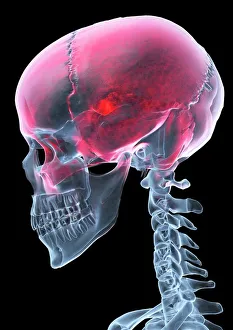
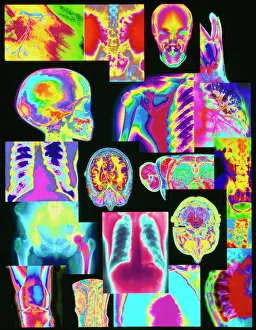
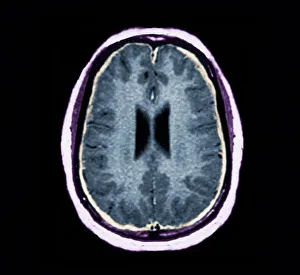
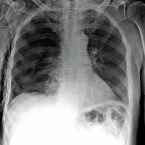
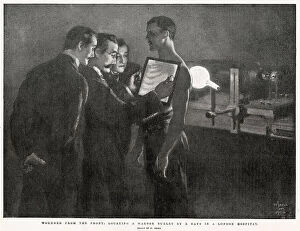
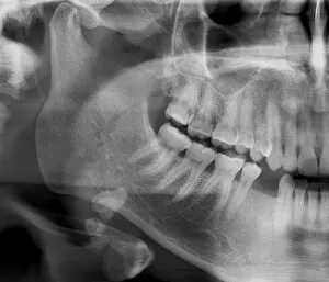
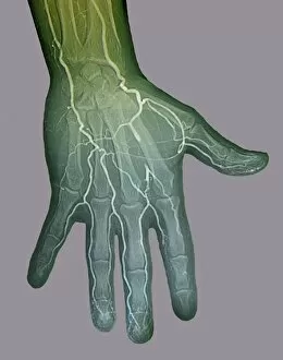
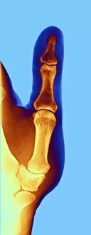
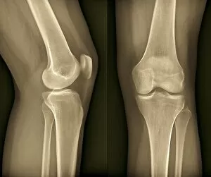
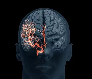
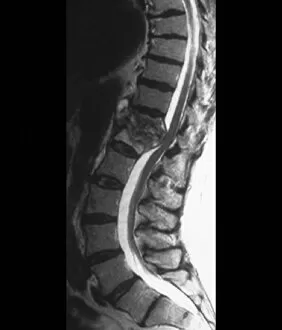
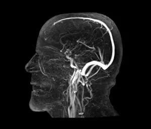
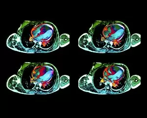
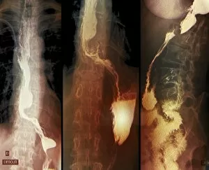
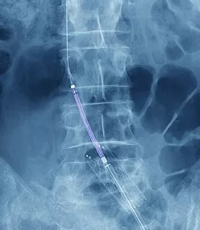
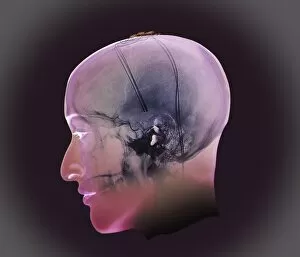
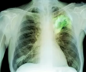
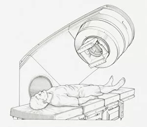
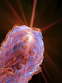
Radiology, the art of peering into the human body's intricate mysteries, reveals a world unseen. From panoramic dental X-rays capturing every tooth's tale to an assortment of coloured X-rays and body scans painting a vibrant picture of our inner workings, this field never ceases to amaze. Intriguingly, even mundane ailments like headaches can transform into captivating X-ray artwork that showcases the hidden source of pain. Hands gracefully suspended in time through an X-ray lens remind us of their tireless work in our daily lives. A healthy knee caught on film reminds us of the marvels within our own bodies. Conversely, tension pneumothorax exposes a fragile balance disrupted by life-threatening conditions that require immediate attention. Picture No. 11676149 captures a moment frozen in time—a snapshot revealing secrets only visible through radiology's watchful eye. Bacterial meningitis unravels its sinister plot under the scrutiny of an MRI scan while total knee replacement unveils newfound mobility through meticulous X-rays. Delving deeper into history, we find ourselves transported to 1900 when soldiers with bullet-bound wounds were examined using early forms of X-ray technology—an extraordinary testament to radiology's role in saving lives during times of conflict. The elegance continues as we witness a healthy ankle joint basking in its uninhibited movement—a reminder that sometimes it is what lies beneath that truly matters. And scoliosis presents itself boldly on an X-ray canvas—each curve telling a unique story etched upon the spine's landscape. Radiology holds within it countless tales waiting to be discovered—one image at a time, and is both science and art intertwined—a window into humanity's most intimate details and profound struggles. With each click and flash, radiologists illuminate paths towards healing and understanding—the guardians who unlock the secrets concealed within our very bones.