
Rbcs Collection
"Exploring the Intricate World of RBCs: Unveiling the Marvels Within Our Blood" Delving into the microscopic realm, we encounter red blood cells (RBCs


All Professionally Made to Order for Quick Shipping
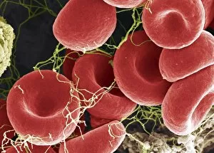
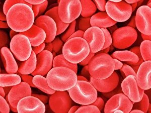
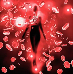
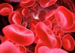
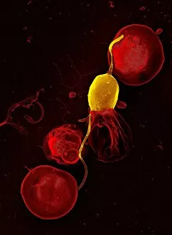
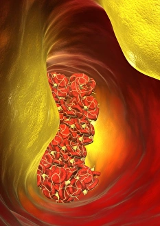
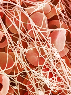
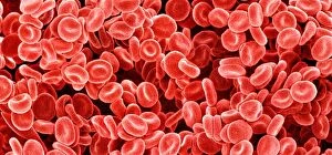
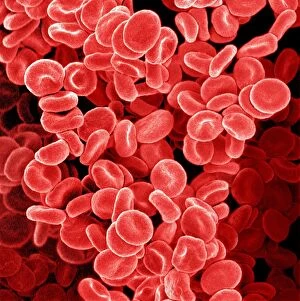
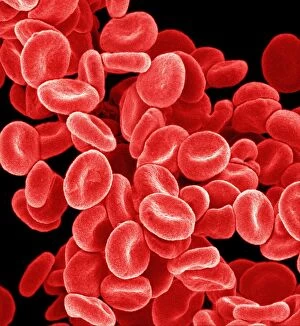
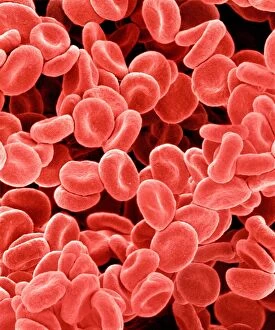
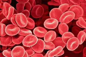
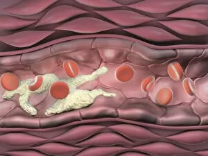
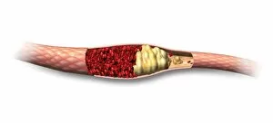
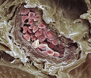
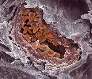
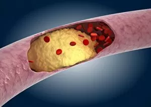
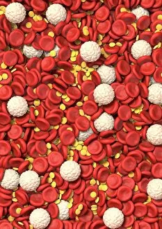
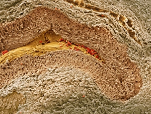
"Exploring the Intricate World of RBCs: Unveiling the Marvels Within Our Blood" Delving into the microscopic realm, we encounter red blood cells (RBCs), the unsung heroes that keep our bodies thriving. Witnessing their beauty under a scanning electron microscope (SEM), we are mesmerized by the intricate structure and delicate nature of these vital blood cells. In a battle against mouse malaria parasites, SEM reveals how RBCs become battlegrounds, showcasing their resilience in defending our immune system. Another captivating SEM image showcases a cluster of healthy red blood cells, highlighting their role in oxygen transportation throughout our body's vast network. Combining science and artistry, computer artwork brings to life vibrant red blood cells pulsating with life force - an awe-inspiring representation of their significance within us. The symbiotic dance between RBCs and our heart is unveiled as they work harmoniously to ensure oxygen-rich blood reaches every corner of our being. A striking SEM image captures the formation of a blood clot - an essential mechanism that prevents excessive bleeding while reminding us of its potential dangers when uncontrolled. Light micrograph C016 / 3035 offers a glimpse into the intricacies of red blood cells at higher magnification, revealing their unique characteristics and diversity within each individual. Through computer artwork depicting a blocked artery, we gain insight into how compromised circulation can impact overall health - emphasizing the importance of maintaining healthy RBC function. Zooming in further with SEM P260 / 0123 exposes fascinating details about blood clots; these intricate formations play crucial roles but require balance for optimal well-being. Crenated red blood cells caught under SEM C016 / 9029 showcase abnormalities that may arise due to various medical conditions or external factors – underscoring why understanding them is paramount for diagnosis and treatment.