Fraud Blocker Skip to main content

Red Blood Cell Collection

Red blood cells, also known as erythrocytes, are the most abundant type of blood cell in our bodies

Background imageRed Blood Cell Collection: Blood cells

Blood cells, computer artwork. Red blood cells (red), or erythrocytes, are biconcave disc-shaped cells that are responsible for supplying tissues with oxygen

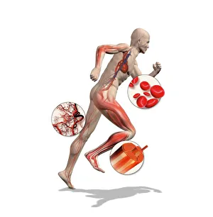
Background imageRed Blood Cell Collection: Athlete physiology, artwork

Athlete physiology, artwork
Athlete physiology. Computer artwork of a male athlete, with enlargements to show the important physiological enhancements typical of most athletes

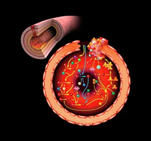
Background imageRed Blood Cell Collection: Blood coagulation cascade, artwork C016 / 9873

Blood coagulation cascade, artwork C016 / 9873
Blood coagulation cascade. Artwork of the biochemical cascade of blood chemicals and proteins during blood clotting (coagulation). The blood vessel and its layered wall is at upper left

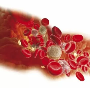
Background imageRed Blood Cell Collection: Diagram showing bloodstream inside a vein, red, white blood cells, platelet

Diagram showing bloodstream inside a vein, red, white blood cells, platelet

Background imageRed Blood Cell Collection: Blood clot, SEM C016 / 9753

Blood clot, SEM C016 / 9753
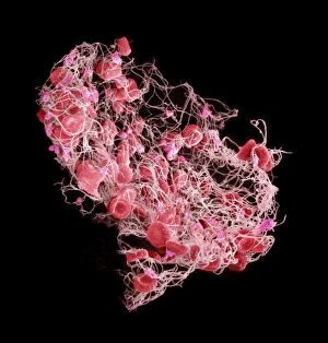
Blood clot. Coloured scanning electron micrograph (SEM) of a blood clot from the inner wall of the left ventricle of a heart

Background imageRed Blood Cell Collection: Blood clot, SEM C016 / 9747

Blood clot, SEM C016 / 9747
Blood clot, coloured scanning electron micrograph (SEM). Red blood cells (erythrocytes) are trapped within a fibrin protein mesh (beige)

Background imageRed Blood Cell Collection: Red blood cells, SEM

Red blood cells, SEM
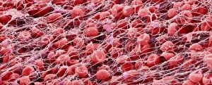
Red blood cells, coloured scanning electron micrograph (SEM)

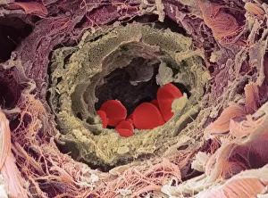
Background imageRed Blood Cell Collection: Blood vessel

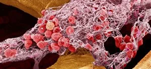
Blood vessel. Coloured scanning electron micrograph (SEM) of a section through a blood vessel in the skin. The vessel contains red blood cells (erythrocytes, red). Magnification unknown

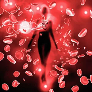
Background imageRed Blood Cell Collection: Red blood cells and heart

Red blood cells and heart. Computer artwork of a heart on the silhouette of a person and red blood cells (erythrocytes). The heart is a hollow muscle that pumps blood around the body

Background imageRed Blood Cell Collection: Blood clot, SEM

Blood clot, SEM
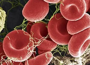
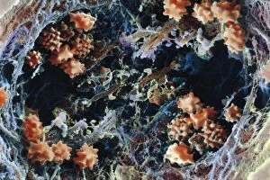
Blood clot, coloured scanning electron micrograph (SEM). Red blood cells are red and fibrin protein strands are green. Platelets are at bottom right

Background imageRed Blood Cell Collection: Red blood cells, SEM

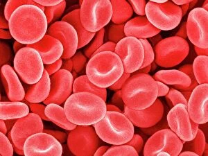
Red blood cells, SEM
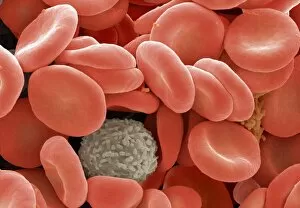
Red blood cells. Coloured scanning electron micrograph (SEM) of red blood cells (erythrocytes). Red blood cells are biconcave, disc-shaped cells that transport oxygen from the lungs to body cells

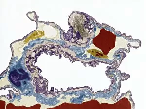
Background imageRed Blood Cell Collection: Lung tissue, TEM

Lung tissue, TEM
Lung tissue, coloured transmission electron micrograph (TEM). A single alveolus is at center. At top right is a pulmonary capillary containing a red blood cell (red) and a platelet (yellow)

Background imageRed Blood Cell Collection: Red blood cells, computer artwork

Red blood cells, computer artwork
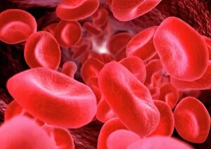
Red blood cells. Computer artwork of human red blood cells (erythrocytes) in a blood vessel

Background imageRed Blood Cell Collection: Red blood cells

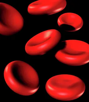
Red blood cells. Computer artwork of human red blood cells (erythrocytes)

Background imageRed Blood Cell Collection: Mouse malaria parasite, SEM

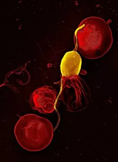
Mouse malaria parasite, SEM
Mouse malaria parasite. Coloured scanning electron micrograph (SEM) of a Plasmodium berghei protozoan (yellow) and red blood cells. P. berghei is the parasite that causes malaria in mice

Background imageRed Blood Cell Collection: A cartoon illustration of a red blood cell looking bored

A cartoon illustration of a red blood cell looking bored

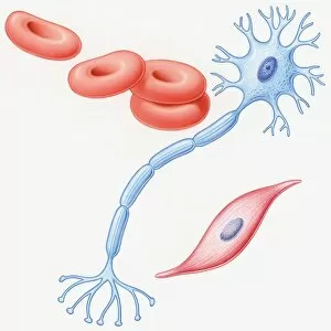
Background imageRed Blood Cell Collection: Illustration of human muscle cell, nerve cell, and red blood cells

Illustration of human muscle cell, nerve cell, and red blood cells

Background imageRed Blood Cell Collection: Coloured SEM of red blood cells, rouleau formation

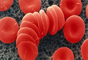
Coloured SEM of red blood cells, rouleau formation
Red blood cells. Coloured scanning electron micrograph of red blood cells in rouleau formation. They often stack together in this way because of their shape

Background imageRed Blood Cell Collection: Red and white blood cells

Red and white blood cells. Coloured scanning electron micrograph (SEM) showing two types of human blood cells

Background imageRed Blood Cell Collection: Diabetes education, blood viscosity

Diabetes education, blood viscosity
MODEL RELEASED. Diabetes education. Nurse using models to demonstrate high blood viscosity to a diabetic patient

Background imageRed Blood Cell Collection: Blood clot, artwork C016 / 4619

Blood clot, artwork C016 / 4619
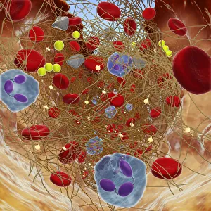
Blood clot in a blood vessel, computer artwork. Red blood cells (erythrocytes) are trapped within a fibrin protein mesh (cream)

Background imageRed Blood Cell Collection: Red blood cells, light micrograph C016 / 3035

Red blood cells, light micrograph C016 / 3035
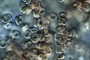
Red blood cells. Differential interference contrast (DIC) micrograph of red blood cells (erythrocytes)

Background imageRed Blood Cell Collection: Red blood cell, SEM

Red blood cell, SEM
Red blood cell, coloured scanning electron micrograph (SEM). Red blood cells (erythrocytes) are carriers of oxygen and carbon dioxide

Background imageRed Blood Cell Collection: False-colour SEM of blood cells on endocardium

False-colour SEM of blood cells on endocardium
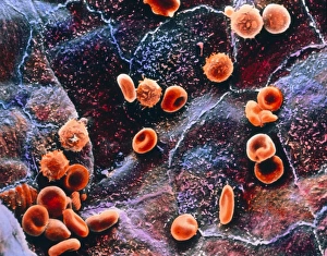
Red Blood cells inside the heart: false-colour scanning electron micrograph (SEM) of red & white blood cells on the endocardium, the lining of the heart

Background imageRed Blood Cell Collection: Cardiac muscle and capillary, TEM

Cardiac muscle and capillary, TEM
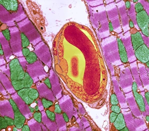
Cardiac muscle. Coloured transmission electron micrograph (TEM) of cardiac muscle fibrils (purple)from a healthy heart. Mitochondria (green) supply the muscle cells with energy

Background imageRed Blood Cell Collection: Blocked artery, computer artwork

Blocked artery, computer artwork
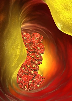
Blocked artery. Computer artwork of a clot of blood cells (thrombus, red) in an artery affected by atherosclerosis. This is a narrowing of an artery due to fatty deposits (yellow) on its inner walls

Background imageRed Blood Cell Collection: Narrowed artery, computer artwork

Narrowed artery, computer artwork
Narrowed artery. Computer artwork of a narrowed artery, showing red blood cells (red) and high levels of low density lipoprotein (LDL, yellow balls)

Background imageRed Blood Cell Collection: Six red blood cells

Six red blood cells

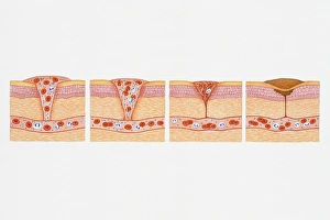
Background imageRed Blood Cell Collection: Illustration of formation of blood clot in series of four cross section illustrations showing blood oozing from blood

Illustration of formation of blood clot in series of four cross section illustrations showing blood oozing from blood vessel through wound before forming platelet plug

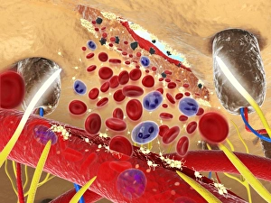
Background imageRed Blood Cell Collection: Digital cross section illustration wound below human skin showing red and white blood cells and yell

Digital cross section illustration wound below human skin showing red and white blood cells and yell

Background imageRed Blood Cell Collection: Cross section showing wound below skin

Cross section showing wound below skin, long fibrin threads trapping red blood, yellow platelets cau

Background imageRed Blood Cell Collection: Illustration of immune response

Illustration of immune response, involving chain of defensive white blood cells, triggered by microb

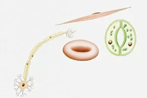
Background imageRed Blood Cell Collection: Illustration of different types of cells

Illustration of different types of cells, nerve cells, red blood cell, muscle cell, and guard cells of a leaf

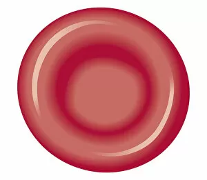
Background imageRed Blood Cell Collection: Biomedical illustration of red blood cell

Biomedical illustration of red blood cell

Background imageRed Blood Cell Collection: Cross section biomedical illustration of red blood cell

Cross section biomedical illustration of red blood cell

Background imageRed Blood Cell Collection: Human Digestive System, Liver, red blood cells under microscope

Human Digestive System, Liver, red blood cells under microscope
Medicine - Human Histology - Digestive system - Liver, red blood cells under microscope

Background imageRed Blood Cell Collection: Circulatory system, Heart, Red blood cells on the endocardium under microscope

Circulatory system, Heart, Red blood cells on the endocardium under microscope
Medicine - Human Histology - Circulatory system - Heart - Red blood cells on the endocardium under microscope

Background imageRed Blood Cell Collection: Cardiac cavity with red blood cells

Cardiac cavity with red blood cells
Medicine - Human Histology - Circulatory system - Heart - Cardiac cavity with red blood cells under the microscope

Background imageRed Blood Cell Collection: Endocardium with red blood cells

Endocardium with red blood cells
Medicine - Human Histology - Circulatory system - Heart - Endocardium with red blood cells under the microscope

Background imageRed Blood Cell Collection: Blood clot, SEM C016 / 9751

Blood clot, SEM C016 / 9751
Blood clot, coloured scanning electron micrograph (SEM). Red blood cells (erythrocytes) are trapped within a fibrin protein mesh (beige)

Background imageRed Blood Cell Collection: Blood clot, SEM C016 / 9746

Blood clot, SEM C016 / 9746
Blood clot, coloured scanning electron micrograph (SEM). Red blood cells (erythrocytes) are trapped within a fibrin protein mesh (beige)

Background imageRed Blood Cell Collection: Blood clot, SEM C016 / 9749

Blood clot, SEM C016 / 9749
Blood clot, coloured scanning electron micrograph (SEM). Red blood cells (erythrocytes) are trapped within a fibrin protein mesh (beige)

Background imageRed Blood Cell Collection: Blood clot, SEM C017 / 7141

Blood clot, SEM C017 / 7141
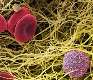
Blood clot, coloured scanning electron micrograph (SEM). Red blood cells (erythrocytes) and a white blood cell (purple) trapped within a fibrin protein mesh (cream)

Background imageRed Blood Cell Collection: Blood clot, SEM C016 / 9750

Blood clot, SEM C016 / 9750
Blood clot, coloured scanning electron micrograph (SEM). Red blood cells (erythrocytes) are trapped within a fibrin protein mesh (beige)

Background imageRed Blood Cell Collection: Blood clot, SEM C016 / 9752

Blood clot, SEM C016 / 9752
Blood clot, coloured scanning electron micrograph (SEM). Red blood cells (erythrocytes) are trapped within a fibrin protein mesh (beige)

Background imageRed Blood Cell Collection: Red blood cells, artwork F006 / 3377

Red blood cells, artwork F006 / 3377
Red blood cells, computer artwork

Background imageRed Blood Cell Collection: West nile virus, artwork F007 / 7822

West nile virus, artwork F007 / 7822
West nile virus, computer artwork

Background imageRed Blood Cell Collection: Dengue virus, artwork F007 / 7818

Dengue virus, artwork F007 / 7818
Dengue virus, computer artwork

Background imageRed Blood Cell Collection: Human rhinovirus 14, artwork F007 / 7852

Human rhinovirus 14, artwork F007 / 7852
Human rhinovirus 14, computer artwork

Background imageRed Blood Cell Collection: Dengue virus, artwork F007 / 7828

Dengue virus, artwork F007 / 7828
Dengue virus, computer artwork

Background imageRed Blood Cell Collection: Sindbis virus, artwork F007 / 7816

Sindbis virus, artwork F007 / 7816
Sindbis virus, computer artwork

Background imageRed Blood Cell Collection: Blood clot, artwork F007 / 6797

Blood clot, artwork F007 / 6797
Blood clot, computer artwork

Background imageRed Blood Cell Collection: Red blood cells, artwork F007 / 6430

Red blood cells, artwork F007 / 6430
Red blood cells, computer artwork

Background imageRed Blood Cell Collection: Swine vesicular disease virus, artwork

Swine vesicular disease virus, artwork
Red blood cells. Computer artwork of red blood cells in a blood vessel

Background imageRed Blood Cell Collection: Foot and Mouth Disease virus, artwork F007 / 7914

Foot and Mouth Disease virus, artwork F007 / 7914
Foot and Mouth Disease virus, computer artwork

Background imageRed Blood Cell Collection: Human rhinovirus 14, artwork F007 / 7813

Human rhinovirus 14, artwork F007 / 7813
Human rhinovirus 14, computer artwork

Background imageRed Blood Cell Collection: Blood clot, artwork F007 / 6795

Blood clot, artwork F007 / 6795
Blood clot, computer artwork

Background imageRed Blood Cell Collection: Small Pox, artwork F007 / 7922

Small Pox, artwork F007 / 7922
Small Pox, computer artwork

Background imageRed Blood Cell Collection: Human arteries, artwork F008 / 1037

Human arteries, artwork F008 / 1037
Human arteries, computer artwork

Background imageRed Blood Cell Collection: Brain stoke, artwork F008 / 0227

Brain stoke, artwork F008 / 0227
Brain stoke, computer artwork

Background imageRed Blood Cell Collection: Human arteries, artwork F008 / 1025

Human arteries, artwork F008 / 1025
Human arteries, computer artwork

Background imageRed Blood Cell Collection: Human arteries, artwork F008 / 1016

Human arteries, artwork F008 / 1016
Human arteries, computer artwork

Background imageRed Blood Cell Collection: Female cardiovascular system, artwork F008 / 1009

Female cardiovascular system, artwork F008 / 1009
Female cardiovascular system, computer artwork

Background imageRed Blood Cell Collection: Human arteries, artwork F008 / 1024

Human arteries, artwork F008 / 1024
Human arteries, computer artwork

Background imageRed Blood Cell Collection: Human femur, artwork F008 / 0486

Human femur, artwork F008 / 0486
Human femur, computer artwork

Background imageRed Blood Cell Collection: Human skin anatomy, artwork F008 / 0368

Human skin anatomy, artwork F008 / 0368
Human skin anatomy, computer artwork

Background imageRed Blood Cell Collection: Human skin anatomy, artwork F008 / 0369

Human skin anatomy, artwork F008 / 0369
Human skin anatomy, computer artwork

Background imageRed Blood Cell Collection: Red blood cells, artwork F008 / 0202

Red blood cells, artwork F008 / 0202
Red blood cells, computer artwork

Background imageRed Blood Cell Collection: Female cardiovascular system, artwork F008 / 1010

Female cardiovascular system, artwork F008 / 1010
Female cardiovascular system, computer artwork

Background imageRed Blood Cell Collection: Human arteries, artwork F008 / 1036

Human arteries, artwork F008 / 1036
Human arteries, computer artwork

Background imageRed Blood Cell Collection: Human bone structure, artwork F008 / 0294

Human bone structure, artwork F008 / 0294
Human bone structure, computer artwork

Background imageRed Blood Cell Collection: Human skin anatomy, artwork F008 / 0203

Human skin anatomy, artwork F008 / 0203
Human skin anatomy, computer artwork

Background imageRed Blood Cell Collection: Human arteries, artwork F008 / 1023

Human arteries, artwork F008 / 1023
Human arteries, computer artwork

Background imageRed Blood Cell Collection: Blood clot, SEM P260 / 0123

Blood clot, SEM P260 / 0123
Blood clot. Coloured scanning electron micrograph (SEM) of a blood clot. The red blood cells (erythrocytes) are trapped in filaments of fibrin protein (pink)

Background imageRed Blood Cell Collection: LM of live human blood cells

LM of live human blood cells
Light micrograph in interference contrast illumination of human blood cells. The majority of the cells seen are red cells, carriers of oxygen throughout the body

Background imageRed Blood Cell Collection: Colour SEM of red & white blood cells

Colour SEM of red & white blood cells
Blood cells. Coloured scanning electron micrograph (SEM) of human blood showing red and white cells. Red blood cells (erythrocytes) have a characteristic biconcave-disc shape and are numerous

Background imageRed Blood Cell Collection: Blood smear, light micrograph F005 / 6090

Blood smear, light micrograph F005 / 6090
Blood smear. Light micrograph showing normal red and white blood cells

Background imageRed Blood Cell Collection: Red blood cells, artwork F005 / 0633

Red blood cells, artwork F005 / 0633
Red blood cells in a blood vessel, computer artwork

Background imageRed Blood Cell Collection: Blood clot, SEM C016 / 9745

Blood clot, SEM C016 / 9745
Blood clot, coloured scanning electron micrograph (SEM). Red blood cells (erythrocytes) are trapped within a fibrin protein mesh (beige)

Background imageRed Blood Cell Collection: Red blood cells, artwork F006 / 2838

Red blood cells, artwork F006 / 2838
Red blood cells, computer artwork

Background imageRed Blood Cell Collection: Red blood cells, artwork F006 / 2731

Red blood cells, artwork F006 / 2731
Red blood cells, computer artwork

Background imageRed Blood Cell Collection: Red blood cells, artwork F006 / 2732

Red blood cells, artwork F006 / 2732
Red blood cells, computer artwork

Background imageRed Blood Cell Collection: Blood stream, artwork F006 / 2729

Blood stream, artwork F006 / 2729
Blood stream, computer artwork


All products are expertly crafted, using premium materials, tailored to your specifications and promptly shipped

Red blood cells, also known as erythrocytes, are the most abundant type of blood cell in our bodies. These tiny cells play a crucial role in maintaining our overall health and well-being. In athlete physiology they can especially important as they carry oxygen from the lungs to every part of the body. This ensures that muscles receive an adequate supply of oxygen during physical activity, enhancing performance and endurance. Artwork depicting the intricate structure of red blood cells showcases their unique shape - biconcave discs without nuclei. This design allows for flexibility and efficient transport through narrow capillaries. The process of blood coagulation cascade is essential for wound healing and preventing excessive bleeding. Artwork illustrating this complex mechanism highlights how red blood cells interact with platelets and clotting factors to form a stable clot, sealing off damaged vessels. Scanning electron microscopy (SEM) images provide detailed views of various aspects related to red blood cells. One such image displays a close-up view of a blood clot formed by these specialized cells (SEM C016 / 9747). Another SEM image reveals infected red blood cells invaded by mouse malaria parasites (SEM). A diagram showcasing the bloodstream inside a vein demonstrates how red and white blood cells along with platelets flow together within our circulatory system. It emphasizes their collective effort in delivering nutrients, removing waste products, and defending against pathogens. Computer artwork beautifully portrays vibrant red blood cells flowing through arteries and veins, emphasizing their vital role in sustaining life throughout our bodies. Lastly, highlighting the connection between red blood cells and heart reminds us that these remarkable microscopic entities work tirelessly alongside our cardiovascular system to ensure proper circulation throughout every organ and tissue.

© Copyright Media Storehouse, All Rights Reserved 2003 - 2024
Facebook     Pinterest     100% Payment Secure     Reviews IO