
Reproduction Pathology Collection
"Exploring the Intricacies of Reproduction Pathology: Unveiling the Hidden Truths" Reproduction is a complex and fascinating process


All Professionally Made to Order for Quick Shipping
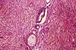
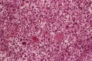
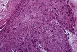
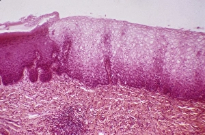
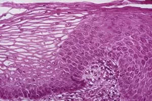
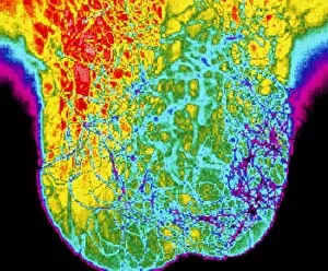
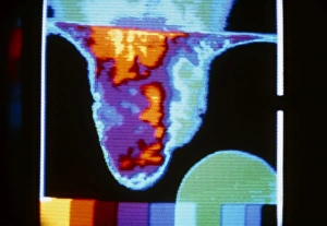
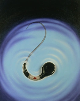
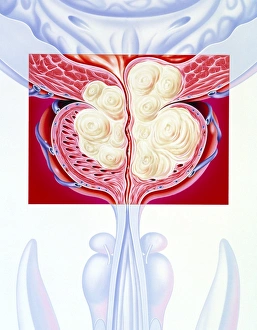
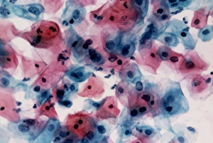
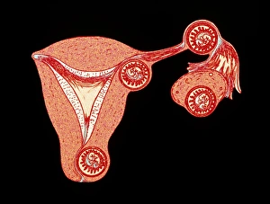
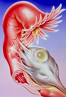
"Exploring the Intricacies of Reproduction Pathology: Unveiling the Hidden Truths" Reproduction is a complex and fascinating process, but sometimes it can be marred by various pathological conditions. In this captivating journey, we delve into the world of reproduction pathology, shedding light on some intriguing aspects that demand our attention. Ovarian cancer, an insidious disease with devastating consequences, takes center stage in our exploration. Through a powerful light micrograph (C015 / 7103), we witness the intricate cellular landscape affected by this formidable condition. The image serves as a poignant reminder of the importance of early detection and intervention. Moving forward, we encounter a deformed sperm cell captured through scanning electron microscopy (SEM). This striking visual representation reminds us that even at the microscopic level, abnormalities can hinder successful fertilization and conception. Our path then leads us to heavy menstrual bleeding depicted through thought-provoking artwork. This portrayal not only highlights its impact on women's lives but also emphasizes the need for effective management strategies to alleviate suffering. Dermoid ovarian cysts make their appearance next - mysterious growths containing tissues from different germ layers. With images like C015 / 6910 and C015 / 6056 showcasing these peculiar formations, we are reminded of nature's enigmatic ways within reproductive organs. The journey continues with cancer of the chorion (C015 / 6905) - an affliction affecting pregnancy-related tissues. Its presence serves as a somber reminder that even during such joyous moments in life, challenges may arise unexpectedly. We then shift our focus towards cervical polyps - abnormal growths within the cervix region. Light micrographs such as C015 / 6744 reveal their intricate structures while endoscope views like C015 / 6738 provide unique perspectives into diagnosis and treatment options for these benign yet bothersome entities.