Fraud Blocker Skip to main content

Reproductive Part Collection (page 4)

"Exploring the Intricate World of Reproductive Parts

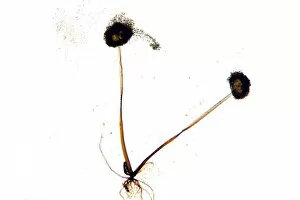
Background imageReproductive Part Collection: Mould fruiting bodies, light micrograph

Mould fruiting bodies, light micrograph. Black mould (Rhizopus nigricans) fruiting bodies (top and upper right) liberating spores

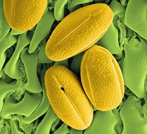
Background imageReproductive Part Collection: Gorse pollen grains, SEM

Gorse pollen grains, SEM
Gorse (Ulex europaeus) pollen grains (yellow), coloured scanning electron micrograph (SEM). Pollen grains are reproductive structures produced by the male parts of flowering plants

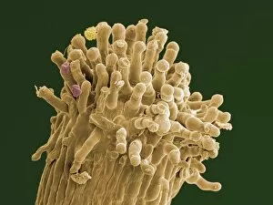
Background imageReproductive Part Collection: Hypericum stigma with pollen grains, SEM

Hypericum stigma with pollen grains, SEM
Hypericum stigma (brown) with pollen grains (pink and yellow), coloured scanning electron micrograph (SEM). Pollen grains are reproductive structures produced by the male parts of flowering plants

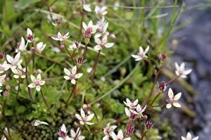
Background imageReproductive Part Collection: Starry saxifrage (Saxifraga stellaris)

Starry saxifrage (Saxifraga stellaris) in flower. Photographed in the Cairngorms National Park, Scottish Highlands, in June

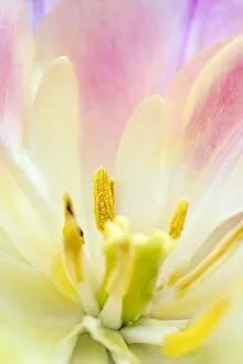
Background imageReproductive Part Collection: Tulip (Tulipa pulchella)

Tulip (Tulipa pulchella) flower. Close-up of the centre of a tulip flower showing the reproductive parts. Six stamens can be seen in a ring around the stigma (pale yellow)

Background imageReproductive Part Collection: Common wasp feeding on a flower

Common wasp feeding on a flower
Common wasp. Close-up of a common wasp (Vespula vulgaris) feeding on nectar from bramble (Rubus sp.) flower. Photographed in August

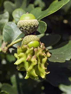
Background imageReproductive Part Collection: Knopper gall on acorn

Knopper gall on acorn. Close-up of two acorns from an Pedunculate Oak (Quercus robur) tree. The lower one has been infected by the parasitic larva of a gall wasp (Andricus quercuscalicis)

Background imageReproductive Part Collection: Knapweed (Centaurea aegialophila)

Knapweed (Centaurea aegialophila)
Knapweed flower (Centaurea aegialophila) on a beach. This flower is endemic to Cyprus

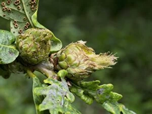
Background imageReproductive Part Collection: Artichoke Galls on Pedunculate Oak

Artichoke Galls on Pedunculate Oak (Quercus rubus). Flower buds infected by the gall wasp Andricus fecundator. In spring the female gall wasp lays single eggs in buds of the Pedunculate oak

Background imageReproductive Part Collection: Scarlet pimpernel flowers

Scarlet pimpernel flowers. The blue form is Anagallis arvensis caerulea and the red form is Anagallis arvensis arvensis. Photographed in Crete

Background imageReproductive Part Collection: Cypriot tulip flower (Tulipa cypria)

Cypriot tulip flower (Tulipa cypria). This plant is endemic to Cyprus. Photographed in the Akamas peninsula in Cyprus

Background imageReproductive Part Collection: Arisaema consanguineum fruit

Arisaema consanguineum fruit
Jack in the pulpit (Arisaema consanguineum). Close-up of the ripening fruit of Arisaema consanguineum. The individual seeds start off green and then gradually turn red as they ripen

Background imageReproductive Part Collection: Fennel seeds, SEM

Fennel seeds, SEM
Fennel (Foeniculum vulgare) seeds, coloured scanning electron micrograph (SEM). As well as its culinary use, Fennel is also commonly used to treat complaints such as indigestion, stomach pains

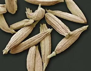
Background imageReproductive Part Collection: Cumin seeds, SEM

Cumin seeds, SEM
Cumin seeds, coloured scanning electron micrograph (SEM). These seeds are from Cuminum cyminum, a flowering plant in the family Apiaceae

Background imageReproductive Part Collection: Ladys mantle reproductive parts, SEM

Ladys mantle reproductive parts, SEM
Ladys mantle reproductive parts, coloured scanning electron micrograph (SEM). Ladys mantle, a plant in the genus Alchemilla

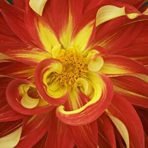
Background imageReproductive Part Collection: Dahlia flower

Dahlia flower. Photographed in the Delta region of British Columbia, Canada

Background imageReproductive Part Collection: Pacific rhododendron

Pacific rhododendron (Rhododendron macrophyllum) flower pistils and stamen, Photographed in Manning Provincial Park, in south-western British Columbia, Canada

Background imageReproductive Part Collection: Nigella seed, SEM

Nigella seed, SEM
Nigella seed. Coloured scanning electron micrograph (SEM) of a seed from a nigella flower (family Ranunculaceae). A seed contains an embryo (a fertilised ovule from which a new plant can grow)

Background imageReproductive Part Collection: Creeping zinnia seed, SEM

Creeping zinnia seed, SEM
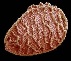
Creeping zinnia seed. Coloured scanning electron micrograph (SEM) of a seed from a creeping zinnia flower (Sanvitalia sp.)

Background imageReproductive Part Collection: Wildflower seed, SEM

Wildflower seed, SEM
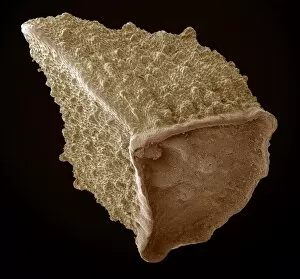
Wildflower seed. Coloured scanning electron micrograph (SEM) of a seed from a wildflower. Wildflower is a general term for flowers growing wild in nature

Background imageReproductive Part Collection: Red campion seed, SEM

Red campion seed, SEM
Red campion seed. Coloured scanning electron micrograph (SEM) of a seed from a red campion flower (Silene dioica). A seed contains an embryo (a fertilised ovule from which a new plant can grow)

Background imageReproductive Part Collection: Copihue seed, SEM

Copihue seed, SEM
Copihue seed. Coloured scanning electron micrograph (SEM) of a seed from a copihue, or Chilean glory, flower (Lapageria rosea)

Background imageReproductive Part Collection: Cactus seed, SEM

Cactus seed, SEM
Cactus seed. Coloured scanning electron micrograph (SEM) of a seed from a cactus flower (family Cactaceae). A seed contains an embryo (a fertilised ovule from which a new plant can grow)

Background imageReproductive Part Collection: Corncockle seed, SEM

Corncockle seed, SEM
Corncockle seed. Coloured scanning electron micrograph (SEM) of a seed from a corncockle flower (Agrostemma githago). A seed contains an embryo (a fertilised ovule from which a new plant can grow)

Background imageReproductive Part Collection: Borage seed, SEM

Borage seed, SEM
Borage seed. Coloured scanning electron micrograph (SEM) of a seed from a borage flower (Borago officinalis). A seed contains an embryo (a fertilised ovule from which a new plant can grow)

Background imageReproductive Part Collection: Anther surface, SEM

Anther surface, SEM
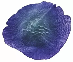
Anther surface. Coloured scanning electron micrograph (SEM) of a close-up of the surface of an anther. An anther is the male reproductive part of a plant

Background imageReproductive Part Collection: Magnolia x wiesneri

Magnolia x wiesneri
Magnolia flower (Magnolia x wiesneri)

Background imageReproductive Part Collection: Camellia flower (Camellia sp. )

Camellia flower (Camellia sp. )
Camellia flower (Camellia sp.)

Background imageReproductive Part Collection: Spider lily (Hymenocallis harrisiana)

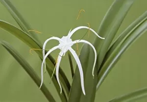
Spider lily (Hymenocallis harrisiana)
Spider lily flower (Hymenocallis harrisiana)

Background imageReproductive Part Collection: Tulipa Peaches and Cream

Tulipa Peaches and Cream
Tulip flower ( Tulipa Peaches and Cream )

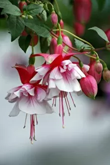
Background imageReproductive Part Collection: Fuchsia Spion Kop

Fuchsia Spion Kop
Fuchsia flowers (Fuchsia Spion Kop ). Photographed in July


All Professionally Made to Order for Quick Shipping

"Exploring the Intricate World of Reproductive Parts: A Microscopic Journey" Embark on a fascinating journey into the hidden realm of reproductive parts as we delve into the captivating beauty and intricate structures of various plant species. From English oak acorns to delicate passion flower pollen, each image captured under a scanning electron microscope (SEM) unveils nature's remarkable mechanisms for reproduction. Witness the mesmerizing patterns and textures present in dandelion pollen grains, resembling tiny spheres adorned with delicate spikes. Philadelphia fleabane pollen grains showcase their unique shape and surface features, while gorse stigma reveals its vibrant yellow hue along with an abundance of attached pollen grains. A striking contrast emerges when lily pollen grain finds its temporary resting place on a rosemary leaf, creating an unexpected juxtaposition between two distinct botanical elements. Forsythia's bright yellow petals give way to its SEM-captured pollen grains that appear like miniature golden orbs suspended in mid-air. Marvel at chickweed's minuscule yet intricately designed pollen grains, showcasing exquisite details that are often overlooked by the naked eye. Passion flower takes center stage once again as its SEM image highlights the complex structure and interwoven threads within each individual grain. Chinese hibiscus surprises us with its vibrant red coloration and distinctive spherical-shaped pollens that seem almost otherworldly under high magnification. Bellflower adds another layer of diversity to our exploration with its own unique set of SEM-captured pollens boasting intriguing shapes and textures. Finally, we encounter thale cress flower micrograph – a stunning portrayal capturing this small flowering plant's reproductive part in all its glory. Delve deeper into this microscopic world where every detail holds significance in ensuring successful reproduction for these remarkable plants. Through these captivating images taken through powerful microscopy techniques, we gain a newfound appreciation for nature's ingenuity in propagating life itself – reminding us how even the tiniest components play a crucial role in the grand tapestry of existence.