Fraud Blocker Skip to main content

Respiratory Collection (page 3)

"The Marvels of the Respiratory System: Exploring the Gateway to Life" Diagram of the Lungs and Bronchial Tubes

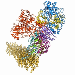
Background imageRespiratory Collection: Oxidoreductase enzyme complex F006 / 9725

Oxidoreductase enzyme complex F006 / 9725
Oxidoreductase enzyme complex, molecular model. This is the membrane-bound domain formed from of a complex of NADH-quinone oxidoreductase subunits

Background imageRespiratory Collection: Oxidoreductase enzyme complex F006 / 9700

Oxidoreductase enzyme complex F006 / 9700
Oxidoreductase enzyme complex. Molecular model of a complex of NADH-quinone oxidoreductase subunits. The whole is termed respiratory complex I

Background imageRespiratory Collection: Lung cancer, artwork

Lung cancer, artwork
Lung cancer, computer artwork

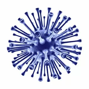
Background imageRespiratory Collection: H1N1 flu virus particle, artwork F006 / 9186

H1N1 flu virus particle, artwork F006 / 9186
H1N1 flu virus particle. Computer artwork of an H1N1 influenza A (flu) virus particle (virion). In the particles lipid envelope (purple) are two types of protein spike

Background imageRespiratory Collection: Reovirus particles, TEM C016 / 9443

Reovirus particles, TEM C016 / 9443
Reovirus particles. Coloured transmission electron micrograph (TEM) of numerous reovirus particles (virions, round). Reoviruses affect the gastrointestinal system and respiratory tract

Background imageRespiratory Collection: Reovirus particles, TEM C016 / 9444

Reovirus particles, TEM C016 / 9444
Reovirus particles. Coloured transmission electron micrograph (TEM) of numerous reovirus particles (virions, blue). Reoviruses affect the gastrointestinal system and respiratory tract

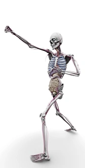
Background imageRespiratory Collection: Male skeleton and organs, artwork

Male skeleton and organs, artwork
Male skeleton and organs and blood vessels, artwork

Background imageRespiratory Collection: Bird flu, conceptual artwork

Bird flu, conceptual artwork
Bird flu. Conceptual artwork of a chicken and flying syringes, representing measures taken against the spread of avian influenza (bird flu)

Background imageRespiratory Collection: Cytochrome c oxidase molecule

Cytochrome c oxidase molecule
Cytochrome c oxidase. Molecular model of a cytochrome c oxidase enzyme from the mitochondria of a cows heart. Cytochrome molecules perform oxidation and reduction reactions for electron transport

Background imageRespiratory Collection: Bird flu research, artwork

Bird flu research, artwork
Bird flu research. Artwork of wild birds tagged with radio transmitters to allow researchers to record and study their migration patterns

Background imageRespiratory Collection: Kidney mitochondria, TEM

Kidney mitochondria, TEM
Kidney mitochondria. Transmission electron micrograph (TEM) of a section through a kidney tubule, showing numerous mitochondria (blue)

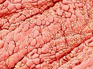
Background imageRespiratory Collection: Trachea lining, SEM C015 / 9939

Trachea lining, SEM C015 / 9939
Trachea lining. Coloured scanning electron micrograph (SEM) of a section through the lining of the trachea (wind pipe). The trachea links the larynx (voice box) to the lungs

Background imageRespiratory Collection: Trachea lining, SEM C015 / 9610

Trachea lining, SEM C015 / 9610
Trachea lining. Coloured scanning electron micrograph (SEM) of the lining of the trachea (wind pipe). The trachea links the larynx to the lungs

Background imageRespiratory Collection: Fish gills, SEM C015 / 8613

Fish gills, SEM C015 / 8613
Fish gills. Coloured scanning electron micrograph (SEM) of filaments from fish gills. These filaments provide a large surface area for the exchange of carbon dioxide for oxygen

Background imageRespiratory Collection: Trachea lining, SEM C015 / 9609

Trachea lining, SEM C015 / 9609
Trachea lining, SEM

Background imageRespiratory Collection: Fish gills, SEM C015 / 8663

Fish gills, SEM C015 / 8663
Fish gills. Coloured scanning electron micrograph (SEM) of filaments from fish gills. These filaments provide a large surface area for the exchange of carbon dioxide for oxygen

Background imageRespiratory Collection: Fish gills, SEM C015 / 8661

Fish gills, SEM C015 / 8661
Fish gills. Coloured scanning electron micrograph (SEM) of filaments from fish gills. These filaments provide a large surface area for the exchange of carbon dioxide for oxygen

Background imageRespiratory Collection: Lung silicosis, light micrograph C015 / 7129

Lung silicosis, light micrograph C015 / 7129
Lung silicosis. Light micrograph of a section through a lung with silicosis, a type of pneumoconiosis caused by exposure to silica dusts. The lung tissue has become fibrous (centre)

Background imageRespiratory Collection: Fish gills, SEM C015 / 8660

Fish gills, SEM C015 / 8660
Fish gills. Coloured scanning electron micrograph (SEM) of filaments from fish gills. These filaments provide a large surface area for the exchange of carbon dioxide for oxygen

Background imageRespiratory Collection: Heart and lung disease, X-rays C018 / 0502

Heart and lung disease, X-rays C018 / 0502
Heart and lung disease. Coloured X-ray (top left) and computed tomography (CT) scans of the chest of an 80 year old patient with heart and lung disease

Background imageRespiratory Collection: Ant spiracle, SEM C018 / 0551

Ant spiracle, SEM C018 / 0551
Ant spiracle, coloured scanning electron micrograph (SEM). Spiracles are openings in an insects exoskeleton that connect to the tracheal tubes which deliver oxygen directly to tissues within

Background imageRespiratory Collection: Jeune syndrome, X-ray C018 / 0410

Jeune syndrome, X-ray C018 / 0410
Jeune syndrome. X-ray of the chest of a 2 year old with Jeune syndrome, also known as asphyxiating thoracic dystrophy. This inherited disease affects the development of the bones and cartilage

Background imageRespiratory Collection: Heart and lung disease, X-ray C018 / 0500

Heart and lung disease, X-ray C018 / 0500
Heart and lung disease. Coloured X-ray of the chest of an 80 year old patient with heart and lung disease. The patient is a former miner and has chronic obstructive pulmonary disease (COPD)

Background imageRespiratory Collection: Jeune syndrome, CT scans C018 / 0411

Jeune syndrome, CT scans C018 / 0411
Jeune syndrome. Coloured computed tomography (CT) scans of the chest of a 2 year old with Jeune syndrome, also known as asphyxiating thoracic dystrophy

Background imageRespiratory Collection: Trachea lining, SEM C016 / 9755

Trachea lining, SEM C016 / 9755
Trachea lining. Coloured scanning electron micrograph (SEM) of the lining of the trachea (wind pipe). The trachea links the larynx to the lungs

Background imageRespiratory Collection: Physical fitness test C014 / 1104

Physical fitness test C014 / 1104
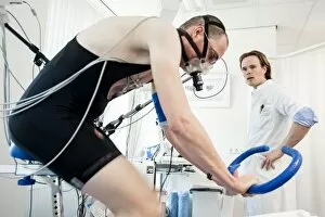
Physical fitness test. Athlete riding an exercise bike while his oxygen consumption is measured with a face mask. Electrodes on his chest monitor his heart rate

Background imageRespiratory Collection: Physical fitness test C014 / 1101

Physical fitness test C014 / 1101
Physical fitness test. Athlete riding an exercise bike while his oxygen consumption is measured with a face mask. Electrodes on his chest monitor his heart rate

Background imageRespiratory Collection: Physical fitness test C014 / 1107

Physical fitness test C014 / 1107
Physical fitness test. Athlete riding an exercise bike while his oxygen consumption is measured with a face mask. Electrodes on his chest monitor his heart rate

Background imageRespiratory Collection: Physical fitness test C014 / 1102

Physical fitness test C014 / 1102
Physical fitness test. Athlete riding an exercise bike while his oxygen consumption is measured with a face mask. Electrodes on his chest monitor his heart rate

Background imageRespiratory Collection: Tuberculosis X-ray diagnosis

Tuberculosis X-ray diagnosis
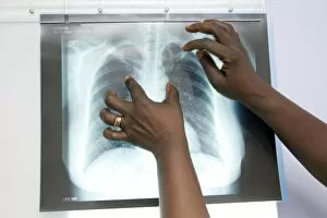
MODEL RELEASED. Tuberculosis (TB) X-ray diagnosis. Close-up of an X-ray of a patients lungs being examined by medical personnel for signs of TB

Background imageRespiratory Collection: Physical fitness test C014 / 1100

Physical fitness test C014 / 1100
Physical fitness test. Athlete undergoing a physical examination. Here his lung capacity is being tested using a spirometer, which measures the volume and speed of air that he can exhale and inhale

Background imageRespiratory Collection: Grape leaf stoma, SEM C014 / 4740

Grape leaf stoma, SEM C014 / 4740
Grape leaf stoma. Coloured scanning electron micrograph (SEM) of a closed stoma (centre-right) on a leaf from a grape (Vitis sp.) vine

Background imageRespiratory Collection: CPR first aid technique, artwork

CPR first aid technique, artwork
CPR first aid technique. Artwork showing the 2nd of 6 steps used in the first aid technique of cardiopulmonary resuscitation (CPR)

Background imageRespiratory Collection: Mitochondrial structure, artwork

Mitochondrial structure, artwork
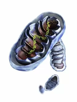
Mitochondrial structure. Computer artwork of a section through a mitochondrion, showing the internal structure and a loop of mitochondrial DNA (deoxyribonucleic acid, mtDNA)

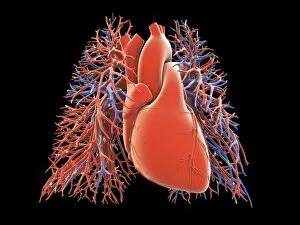
Background imageRespiratory Collection: Heart-lungs circulatory system, artwork

Heart-lungs circulatory system, artwork
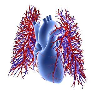
Heart-lungs circulatory system, computer artwork. The heart (centre) is a hollow muscle that pumps blood around the body. The two branches of the pulmonary artery (red)

Background imageRespiratory Collection: Heart-lung circulatory system, artwork

Heart-lung circulatory system, artwork
Heart-lung circulatory system, computer artwork. The heart (centre) is a hollow muscle that pumps blood around the body. The two branches of the pulmonary artery (red)

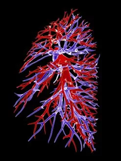
Background imageRespiratory Collection: Lung blood vessel, artwork

Lung blood vessel, artwork
Lung blood vessels, computer artwork. Shown are the lung blood vessels in side view. The branches of the pulmonary artery (red) which carry deoxygenated blood from the heart to the lungs

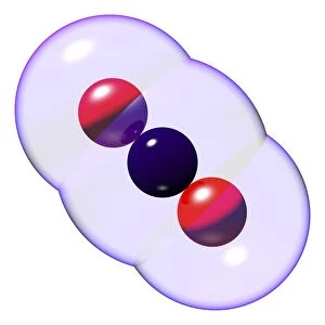
Background imageRespiratory Collection: Carbon dioxide molecules C016 / 8495

Carbon dioxide molecules C016 / 8495
Carbon dioxide molecules. Computer artwork showing the structure of a molecule of carbon dioxide. Carbon dioxide is a colourless gas that occurs naturally in the atmosphere

Background imageRespiratory Collection: Carbon dioxide molecule C016 / 8494

Carbon dioxide molecule C016 / 8494
Carbon dioxide molecule. Computer artwork showing the structure of a molecule of carbon dioxide. Carbon dioxide is a colourless gas that occurs naturally in the atmosphere

Background imageRespiratory Collection: Carbon dioxide molecule C016 / 8493

Carbon dioxide molecule C016 / 8493
Carbon dioxide molecule. Computer artwork showing the structure of a molecule of carbon dioxide. Carbon dioxide is a colourless gas that occurs naturally in the atmosphere

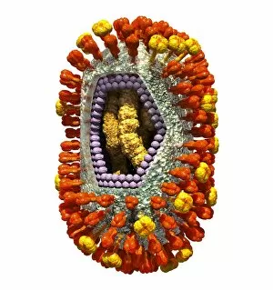
Background imageRespiratory Collection: Influenza virus, artwork C016 / 8345

Influenza virus, artwork C016 / 8345
This image may not be used in educational posters Influenza virus. Cut-away computer artwork of an influenza (flu) virus particle (virion)

Background imageRespiratory Collection: Influenza virus particle, TEM

Influenza virus particle, TEM
Influenza virus particle, coloured transmission electron micrograph (TEM). The virus consists of ribonucleic acid (RNA), surrounded by a nucleocapsid (light green) and a lipid envelope (dark green)

Background imageRespiratory Collection: Heart-lungs circulatory system

Heart-lungs circulatory system, computer artwork. The heart (centre) is a hollow muscle that pumps blood around the body. The two branches of the pulmonary artery (red)

Background imageRespiratory Collection: RSV virus fusion glycoprotein C015 / 6689

RSV virus fusion glycoprotein C015 / 6689
RSV virus fusion glycoprotein, molecular model. This is a fusion glycoprotein of human respiratory syncytial virus (RSV). This glycoprotein is used by the virus to invade host cells

Background imageRespiratory Collection: RSV virus fusion glycoprotein C015 / 6688

RSV virus fusion glycoprotein C015 / 6688
RSV virus fusion glycoprotein, molecular model. This is a fusion glycoprotein of human respiratory syncytial virus (RSV). This glycoprotein is used by the virus to invade host cells

Background imageRespiratory Collection: Oxidoreductase enzyme complex C015 / 6554

Oxidoreductase enzyme complex C015 / 6554
Oxidoreductase enzyme complex, molecular model. This is the membrane-bound domain formed from of a complex of NADH-quinone oxidoreductase subunits

Background imageRespiratory Collection: Smog from wildfires, Tomsk C015 / 5889

Smog from wildfires, Tomsk C015 / 5889
Smog. View of the city of Tomsk, Russia, through a heavy smog caused by local wild fires. Photographed in July 2012

Background imageRespiratory Collection: Oxidoreductase enzyme complex C015 / 6553

Oxidoreductase enzyme complex C015 / 6553
Oxidoreductase enzyme complex, molecular model. This is the membrane-bound domain formed from of a complex of NADH-quinone oxidoreductase subunits


All Professionally Made to Order for Quick Shipping

"The Marvels of the Respiratory System: Exploring the Gateway to Life" Diagram of the Lungs and Bronchial Tubes: Unveiling the intricate network within our bodies that facilitates every breath we take, this diagram showcases the lungs and bronchial tubes, highlighting their vital role in oxygenating our bloodstream. Diagram of the Heart, Lungs, and Windpipe: Witnessing a symphony of life-sustaining organs working harmoniously together, this diagram depicts the heart pumping blood while being surrounded by the lungs and windpipe – a testament to our body's remarkable respiratory system. Illustration of Human Respiratory System: Embark on an anatomical journey through our respiratory system as this illustration reveals its various components such as oral cavity, nasal cavity, larynx, trachea, bronchus, and lungs – each playing a crucial part in ensuring efficient respiration. Tension Pneumothorax X-ray: Peering into medical imaging technology's window into diagnosis and treatment strategies for collapsed lungs caused by tension pneumothorax - an urgent condition requiring immediate attention to restore normal breathing patterns. English Oak Leaf Pores SEM Image: Delving into nature's wonders at microscopic levels reveals mesmerizing images showcasing English oak leaf pores under scanning electron microscopy (SEM), reminding us how even plants have their unique respiratory systems. Cystic Fibrosis: Unmasking a Genetic Challenge Exploring cystic fibrosis - a genetic disorder affecting lung function - sheds light on ongoing research efforts aimed at improving treatments for individuals facing challenges with their respiratory health from an early age. Konstantin Buteyko: Soviet Doctor Revolutionizing Breathing Techniques Discovering how Dr. Konstantin Buteyko revolutionized breathing techniques offers insights into his innovative methods that have helped countless individuals manage asthma symptoms and enhance overall respiratory well-being. Paramyxovirus Particles TEM Image.