Fraud Blocker Skip to main content

Retina Collection

The retina, a fascinating structure within the human eye, has captivated scientists and artists alike throughout history

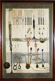
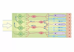
Background imageRetina Collection: Histological Diagram of a Mammalian Retina

Histological Diagram of a Mammalian Retina
RAMON Y CAJAL, Santiago (1852-1934). Spanish doctor and histologist, Nobel Prize in 1906. Histological diagram of a mammalian retina. Original drawing by Ramon y Cajal

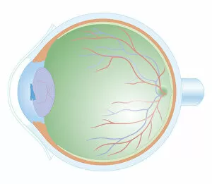
Background imageRetina Collection: Eye anatomy, artwork

Eye anatomy, artwork
Eye anatomy, computer artwork. At the front of the eye is the cornea, a transparent coating. Behind this is the lens, which is partly covered by the iris

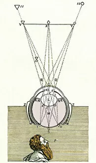
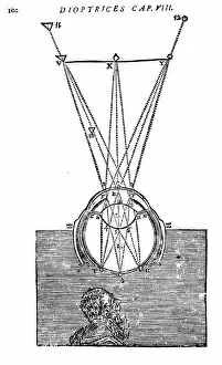
Background imageRetina Collection: Descartes optics theory, 17th century

Descartes optics theory, 17th century
Descartes optics theory, artwork. This is the eye diagram from Descartes book Optics (1637). Three external points are shown depicted on the retina at the back of the eye

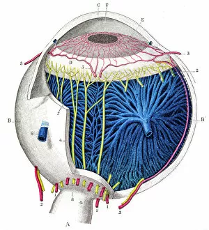
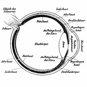
Background imageRetina Collection: Human eye anatomy engraving 1899

Human eye anatomy engraving 1899
Corso Elementare di Scienze Naturali

Background imageRetina Collection: Rod and cone cells of the eye, SEM C014 / 4866

Rod and cone cells of the eye, SEM C014 / 4866
Rod and cone cells of the eye. Coloured scanning electron micrograph (SEM) of rod and cone cells in the retina of a mammalian eye

Background imageRetina Collection: Biometric scans

Biometric scans. Computer artwork of a human eye and fingerprint. Biometrics is the identification of individuals by measuring and analysing their physiological characteristics, such as fingerprints
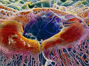
Background imageRetina Collection: False-colour SEM of retina featuring central fovea

False-colour SEM of retina featuring central fovea
Foveal retina. False-colour scanning electron micrograph of the human retina featuring the central fovea, a crater-like depression in the photosensitive layer of the eye

Background imageRetina Collection: Artificial intelligence, conceptual art

Artificial intelligence, conceptual artwork. Electronic circuitry is seen behind the eye

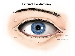
Background imageRetina Collection: External anatomy of the human eye (with labels)

External anatomy of the human eye (with labels)

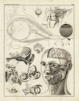
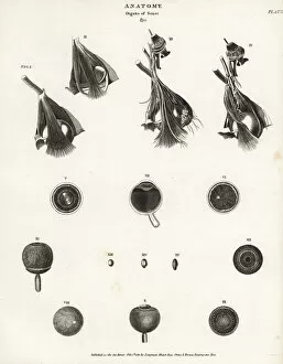
Background imageRetina Collection: Anatomy of the human ear and eye, 1839 (lithograph)

Anatomy of the human ear and eye, 1839 (lithograph)
FLO4684845 Anatomy of the human ear and eye, 1839 (lithograph); (add.info.: Anatomy of the human ear and eye. Lithograph from Lorenz Oken's Universal Natural History)

Background imageRetina Collection: Digital illustration of human eye transforming images made from light into electrical impulses

Digital illustration of human eye transforming images made from light into electrical impulses
arrow sign, biomedical illustration, blue background, connection, cornea, cross section, digitally generated, direction, eyesight, full frame, horizontal, human eye, iris, lens, no people

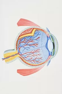
Background imageRetina Collection: Normal anatomy of the eye in cross section

Normal anatomy of the eye in cross section
Horizontal, Vision, Detail, Anatomy, Diagram, Muscle, Illustration, Lens, Iris, Medical, Eyeball, Biology, Lateral, Cut Out, White Background, Eye, Artwork, Cross Section, Color Image, Close Up

Background imageRetina Collection: The eye

The eye
Abbas Arabzadeh

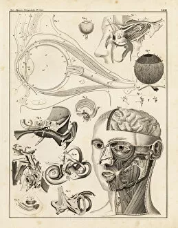
Background imageRetina Collection: Anatomy of the human ear and eye

Anatomy of the human ear and eye. Lithograph from Lorenz Okens Universal Natural History, Allgemeine Naturgeschichte fur alle Stande, Stuttgart, 1839

Background imageRetina Collection: The eye seen through a microscope, from Atlas of Ophthalmoscopy

The eye seen through a microscope, from Atlas of Ophthalmoscopy
CHT236313 The eye seen through a microscope, from Atlas of Ophthalmoscopy, published in Paris, 1870 (chromolitho) by Liebreich

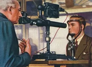
Background imageRetina Collection: Eye-prints, 1938

Eye-prints, 1938. Churchmans cigarette card, from a series titled Modern Wonders [WA & AC Churchman, Great Britain & Ireland, 1938]

Background imageRetina Collection: Descartes explanation of vision, 1692

Descartes explanation of vision, 1692. Light rays being passed through the eye, being focused by the lens (I) and forming images T, S, R on the retina

Background imageRetina Collection: Visual system, Eye, Retina under microscope

Visual system, Eye, Retina under microscope
Medicine - Anatomy - Visual system - Eye - Retina under microscope

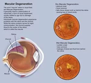
Background imageRetina Collection: Retina with macular degeneration

Retina with macular degeneration

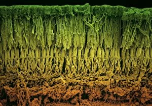
Background imageRetina Collection: Retina layers, SEM C018 / 0520

Retina layers, SEM C018 / 0520
Retina layers, coloured scanning electron micrograph (SEM). The retina of the human eye is found on the inside of the eyeball and is formed from a number of layers

Background imageRetina Collection: Rod and cone cells of the eye, SEM C014 / 4864

Rod and cone cells of the eye, SEM C014 / 4864
Rod and cone cells of the eye. Coloured scanning electron micrograph (SEM) of rod and cone cells in the retina of a mammalian eye

Background imageRetina Collection: Eye, rods and cones of retina, artwork C017 / 7791

Eye, rods and cones of retina, artwork C017 / 7791
Photograph of an human eye and illustration of rods and cones in the retina. The retina, inside the eye, contains a light-sensitive membranous layer of cells

Background imageRetina Collection: SEM of rod & cone cell of the retina

SEM of rod & cone cell of the retina
False-colour scanning electron micrograph (SEM) of rod and cone cells of the eye retina. Cone cells (blue) and more numerous rod cells (pink, purple) are specialized light-sensitive cells

Background imageRetina Collection: Retinal implant, artwork

Retinal implant, artwork
Retinal implant. Computer artwork of a biomedical chip (square, centre right) implanted just under the surface of the retina in a human eye

Background imageRetina Collection: The Ring Magnet

The Ring Magnet
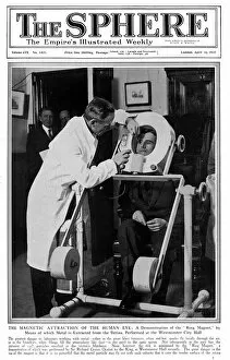
The front cover of " The Sphere" magazine, 16 April 1927, featuring the latest innovation in eye-care. Factory workers suffering from metal filings flicked in to their eye can now have them

Background imageRetina Collection: Anatomy of the human eye

Anatomy of the human eye.. Anatomy of the human eye, showing eyeball, optic nerve, muscle, cornea, retina, sections through eye

Background imageRetina Collection: Human eye, published in 1898

Human eye, published in 1898
Antique illustration of a Human eye, published in 1898

Background imageRetina Collection: Cross-section diagram of human eye

Cross-section diagram of human eye

Background imageRetina Collection: Cross section of human eye

Cross section of human eye

Background imageRetina Collection: Digital illustration of human brain and mapping of visual field onto retina matching arrangement

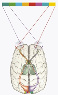
Digital illustration of human brain and mapping of visual field onto retina matching arrangement of data on surface of visual cortex

Background imageRetina Collection: Cross section digital illustration of retina with futuristic retinal implant

Cross section digital illustration of retina with futuristic retinal implant

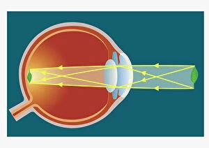
Background imageRetina Collection: Illustration of vision of the human eye

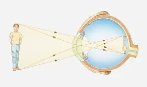
Illustration of vision of the human eye

Background imageRetina Collection: Cross section biomedical illustration of laser surgery for retinopathy

Cross section biomedical illustration of laser surgery for retinopathy

Background imageRetina Collection: Cross section biomedical illustration of how the retina responds to light using nerve cells

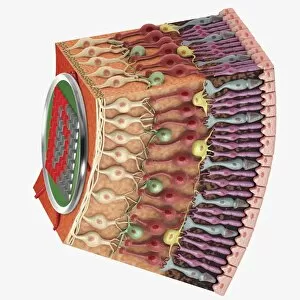
Cross section biomedical illustration of how the retina responds to light using nerve cells, rods and cones

Background imageRetina Collection: Cross section biomedical illustration of anatomy of human eye

Cross section biomedical illustration of anatomy of human eye

Background imageRetina Collection: Anatomy of human eye, perspective

Anatomy of human eye, perspective

Background imageRetina Collection: Orbital cut showing abducent nerve with ciliary ganglion and oculomotor nerve

Orbital cut showing abducent nerve with ciliary ganglion and oculomotor nerve

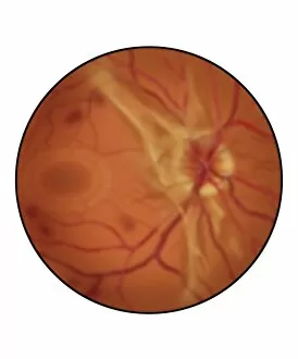
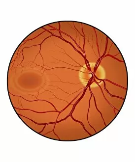
Background imageRetina Collection: Retina of a normal eye

Retina of a normal eye

Background imageRetina Collection: Retina with advanced diabetic retinopathy

Retina with advanced diabetic retinopathy, showing increased scar tissue, hemorrhaging and diffuse vitreous blood (blurred appearance)

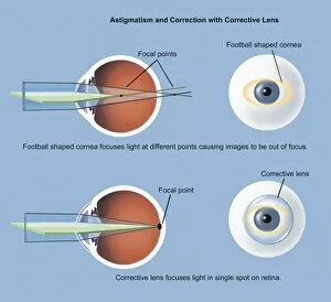
Background imageRetina Collection: Astigmatism and correction with corrective lens

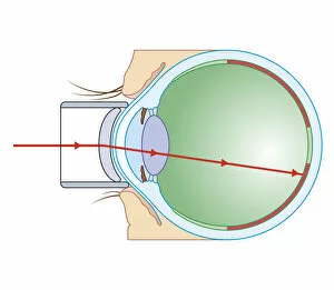
Astigmatism and correction with corrective lens

Background imageRetina Collection: Conceptual image of human eye cross section

Conceptual image of human eye cross section

Background imageRetina Collection: Anatomy of human eye

Anatomy of human eye

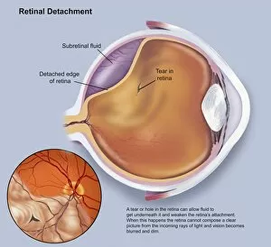
Background imageRetina Collection: Diagram of a retinal detachment

Diagram of a retinal detachment

Background imageRetina Collection: Retina of eye with glaucoma

Retina of eye with glaucoma


All Professionally Made to Order for Quick Shipping

The retina, a fascinating structure within the human eye, has captivated scientists and artists alike throughout history. From histological diagrams to intricate artwork, the intricacies of its anatomy have been meticulously studied and depicted. In the 17th century, Descartes' optics theory shed light on how light interacts with the retina. This groundbreaking understanding paved the way for further exploration into this complex organ. An engraving from 1899 beautifully illustrates the external anatomy of the human eye, showcasing how it works in harmony with the retina. The rod and cone cells of the eye are highlighted in another image taken through a scanning electron microscope (SEM), revealing their delicate structures responsible for vision. Advancements in technology have allowed us to delve deeper into studying retinas through biometric scans. These scans provide valuable insights into individual characteristics and contribute to fields like artificial intelligence. A captivating false-color SEM image showcases a central fovea within a retina—an area crucial for sharp vision—highlighting its intricate details that make it an essential part of our visual system. At a molecular level, rhodopsin protein molecules play a vital role in converting light signals received by retinal cells into electrical impulses sent to our brain—a process critical for sight perception. As we explore further, we discover not only its anatomical features but also its network of blood vessels that supply oxygen and nutrients to sustain this remarkable organ's functionality. From ancient theories to modern scientific discoveries, artful depictions or microscopic examinations—the study of retinas continues to intrigue us as we unravel more about one of nature's most incredible creations: our eyes.