
Rod Shaped Collection
"Exploring the World of Rod-Shaped Bacteria: A Fascinating Microscopic Journey" In the vast realm of microbiology


All Professionally Made to Order for Quick Shipping
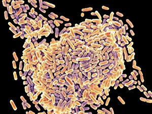
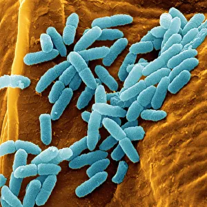
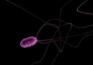
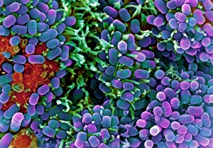
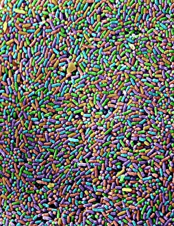
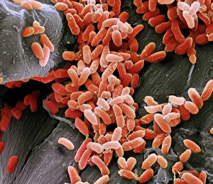
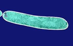
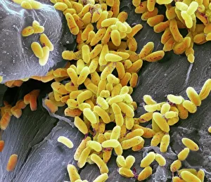
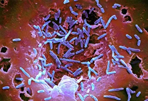
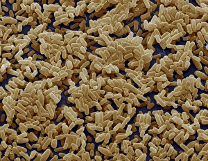
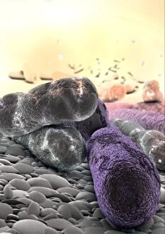
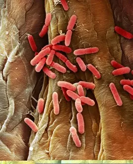
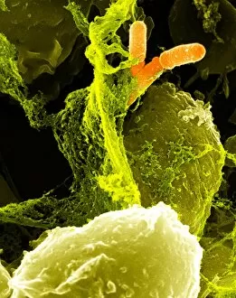
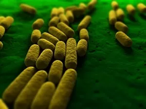
"Exploring the World of Rod-Shaped Bacteria: A Fascinating Microscopic Journey" In the vast realm of microbiology, rod-shaped bacteria have captivated scientists with their unique structures and intriguing characteristics. Through the lens of scanning electron microscopy (SEM) and transmission electron microscopy (TEM), these tiny organisms reveal a mesmerizing world that is both beautiful and complex. One such example is Salmonella bacteria, which appear as slender rods under SEM. These notorious pathogens are responsible for causing foodborne illnesses in humans, reminding us of the importance of proper hygiene practices in our daily lives. Moving on to tuberculosis bacteria, another rod-shaped microbe that can be observed through SEM. This bacterium has plagued humanity for centuries, highlighting the ongoing battle against infectious diseases worldwide. Flagellate bacteria showcase an extraordinary feature – whip-like appendages called flagella that enable them to move with remarkable agility. Their elongated bodies give them a distinctive appearance when examined using SEM. Pseudomonas aeruginosa bacteria exhibit a fascinating morphology when viewed under SEM. With their long rods and polar flagella, they possess incredible adaptability and resilience in various environments. E. coli bacterium reveals its characteristic shape through TEM imaging. This versatile organism exists naturally in our intestines but certain strains can cause severe gastrointestinal infections if ingested improperly cooked food or contaminated water. Mycobacterium chelonae bacteria display their rod-like forms under SEM examination. These environmental microbes are known for causing opportunistic infections primarily affecting individuals with compromised immune systems. Bacteriophages add an artistic touch to this microscopic journey - captivating artworks depicting these viruses that specifically target bacterial cells offer hope for future medical advancements in combating antibiotic-resistant strains effectively. Lastly, we encounter Salmonella typhimurium bacteria once again; however, this time captured beautifully by SEM photography. The intricate details revealed by this technique emphasize the need for continued research into preventing salmonellosis, a common foodborne illness.