Fraud Blocker Skip to main content

Scanning Electron Microscope Collection (page 5)

"Unveiling the Hidden World

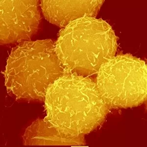
Background imageScanning Electron Microscope Collection: Haematopoietic stem cells, SEM C013 / 5009

Haematopoietic stem cells, SEM C013 / 5009
Haematopoietic stem cells, coloured scanning electron micrograph (SEM). Stem cells can differentiate into any other cell type

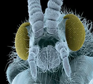
Background imageScanning Electron Microscope Collection: Fly head, colored scanning electron micrograph

Fly head, colored scanning electron micrograph
Fly head, coloured scanning electron micrograph (SEM). Close-up of the head of a fly, showing its short antennae (upper centre), which are seen between its compound eyes (brown)

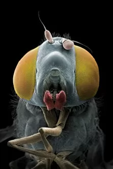
Background imageScanning Electron Microscope Collection: Irresistible

Irresistible
Coloured scanning electron micrograph (SEM) of a hoverfly, also called flower fly or syrphid fly (family Syrphidae). The compound eyes are composed of numerous light sensing organs (ommatidia)

Background imageScanning Electron Microscope Collection: Rust fungus infection, SEM

Rust fungus infection, SEM
Rust fungus infection. Coloured scanning electron micrograph (SEM) of spore-bearing aecia from a rust (Puccinia lagenophora) fungus, on a common groundsel (Senecio vulgaris) plant

Background imageScanning Electron Microscope Collection: Ant compound eye, SEM C018 / 0552

Ant compound eye, SEM C018 / 0552
Ant compound eye, coloured scanning electron micrograph (SEM). The units in a compound eye, the ommatidia, each contain a cornea and photoreceptor cells

Background imageScanning Electron Microscope Collection: Yeast fungus skin infection, SEM

Yeast fungus skin infection, SEM
Yeast fungus skin infection. Coloured scanning electron micrograph (SEM) of human skin infected with a yeast fungus. Magnification: x3130 when printed at 10 centimetres wide

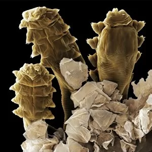
Background imageScanning Electron Microscope Collection: Follicle mite heads (SEM) C013 / 5120

Follicle mite heads (SEM) C013 / 5120
Follicle mite heads (Demodex folliculorum), coloured scanning electron micrograph (SEM) of follicle or eyelash mites protruding from a dissected human hair follicle

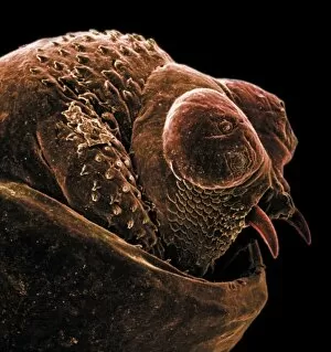
Background imageScanning Electron Microscope Collection: The anus of a bot fly

The anus of a bot fly
Scanning electron microscope image of the anus of a bot fly. Image on display in the Darwin Centre at the Natural History Museum, London

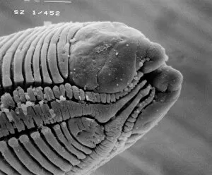
Background imageScanning Electron Microscope Collection: Roundworm

Roundworm
Scanning electron microscope (SEM) image of a parasitic roundworms head (x 1000 on a standard 9 cm wide print)

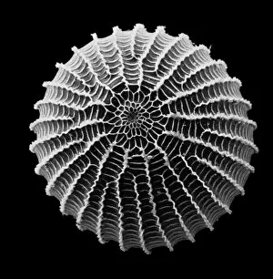
Background imageScanning Electron Microscope Collection: Moth egg

Moth egg
Scanning electron microscope (SEM) image of a moth egg (x 90). The caterpillar emerges by chewing through the shell

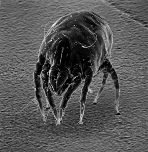
Background imageScanning Electron Microscope Collection: Dermatophagoides sp. dust mite

Dermatophagoides sp. dust mite
Scanning electron microscope image of a dust mite. Dust mites are secondary to pollen as a cause for allergies and they live in bedding, soft furniture and carpets

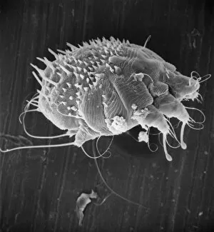
Background imageScanning Electron Microscope Collection: Sarcoptes scabiei, scabies mite

Sarcoptes scabiei, scabies mite
Scanning electron microscope image of an itch or scabies mite, a parasite that infests a wide variety of mammalian hosts including man

Background imageScanning Electron Microscope Collection: Shed claw from a cat, SEM

Shed claw from a cat, SEM
Cats claw, coloured scanning electron micrograph (SEM). This is the shed outer layer of the claw. Domestic cats typically shed the outer layers of their claws, which grow from the inside

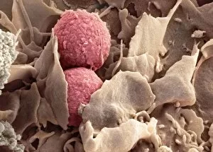
Background imageScanning Electron Microscope Collection: Phagocytosis of fungus spores, SEM

Phagocytosis of fungus spores, SEM
Phagocytosis of fungus spores. Coloured scanning electron micrograph (SEM) of spores from the fungus Aspergillus fumigatus (red) being phagocytosed (engulfed and destroyed) by a dendritic cell (brown)

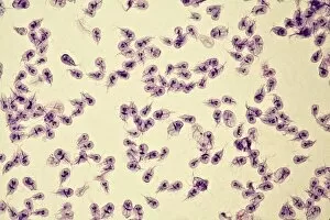
Background imageScanning Electron Microscope Collection: Giardia lamblia protozoa, micrograph

Giardia lamblia protozoa, micrograph
Giardia lamblia protozoa, light micrograph. These single-celled parasitic protozoa infect the intestinal tract and are most common in tropical regions

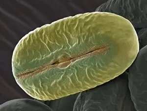
Background imageScanning Electron Microscope Collection: Broad bean pollen, SEM

Broad bean pollen, SEM
Broad bean pollen. Coloured scanning electron micrograph (SEM) of pollen grains from a Vicia faba plant. Magnification: x2000 when printed 10 centimetres wide

Background imageScanning Electron Microscope Collection: Deformed sperm cell, SEM

Deformed sperm cell, SEM
Deformed sperm cell. Coloured scanning electron micrograph (SEM) of double-tailed sperm cell, or spermatozoon. A sperm cells motility may be seriously affected by having multiple heavy tails

Background imageScanning Electron Microscope Collection: Snake head, SEM

Snake head, SEM
Snake (suborder Serpentes) head, coloured scanning electron micrograph (SEM). The snakes forked tongue (right) is used to detect prey by picking up scent particles from the air

Background imageScanning Electron Microscope Collection: Chloroplast, SEM

Chloroplast, SEM
Chloroplast. Coloured scanning electron micrograph (SEM) of a section through a plant cell, showing a fractured chloroplast (green)


All Professionally Made to Order for Quick Shipping

"Unveiling the Hidden World: Exploring with a Scanning Electron Microscope" The scanning electron microscope (SEM) is an incredible tool that allows us to delve into the microscopic realm, revealing intricate details and unlocking secrets of the unseen. With its high magnification capabilities, it offers a glimpse into fascinating aspects of life and nature. In one captivating image, we witness the beauty of gender determination as X and Y chromosomes are captured under the SEM's lens. The intricate patterns and structures within these chromosomes showcase the complexity of our genetic makeup. Moving on to another astonishing discovery, a Scanning Electron Micrograph presents us with an up-close view of a Praying Mantis at 30 times magnification. Every tiny detail becomes visible, from its delicate limbs to its mesmerizing eyes – reminding us of nature's remarkable diversity. Shifting focus to plant life, lavender pollen grains come alive in another SEM image. These minuscule particles take center stage as their unique shapes and textures become apparent under intense scrutiny. Zooming in even further reveals a Tardigrade or 'Water Bear' at an astounding 1250 times magnification. This resilient creature captures our imagination as we explore its alien-like features through this powerful imaging technique. Delving deeper into biological wonders, T lymphocytes battling cancer cells are showcased in yet another breathtaking SEM capture. This visual representation highlights the ongoing fight within our bodies against diseases like cancer – emphasizing both hope and resilience. Calcareous phytoplankton takes center stage next as we uncover their intricate forms through SEM imagery. These microscopic organisms play crucial roles in marine ecosystems while leaving us awe-inspired by their stunningly detailed structures. Transitioning to neural marvels, nerve cells reveal their intricacy under the watchful eye of an SEM. Their branching extensions create complex networks that enable communication throughout our bodies – showcasing nature's ingenuity at work. Venturing into insect realms once again, a fruit fly is immortalized under the SEM's lens.